Article

Variations in the Phytoremediation Efficiency of Metal-polluted Water with Salvinia biloba: Prospects and Toxicological Impacts

by
Julia Emiliani
1,†,
Wendi G. Llatance Oyarce
1,2,†,
C. Daniela Bergara
1,
Lucas M. Salvatierra
1,3,
Luís A. B. Novo
4,* and
Leonardo M. Pérez
1,3,*
1
Grupo de Biotecnología de Materiales y Medioambiente (Bio&TecMA), Instituto de Investigaciones en Ingeniería Ambiental, Química y Biotecnología Aplicada (INGEBIO-UCA), Facultad de Química e Ingeniería del Rosario, Pontificia Universidad Católica Argentina (UCA), Av. Pellegrini 3314, S2002QEO Rosario, Argentina
2
Escuela Profesional de Ingeniería Ambiental, Facultad de Ingeniería Civil y Ambiental, Universidad Nacional Toribio Rodríguez de Mendoza de Amazonas (UNTRM), 01001 Chachapoyas, Peru
3
National Council for Scientific and Technical Research (CONICET), Ministry of Science, Technology and Productive Innovation, Godoy Cruz 2290, C1425FQB Buenos Aires, Argentina
4
Laboratory of Experimental and Applied Phytotechnologies (LEAPH), GeoBioTec, Department of Geosciences, University of Aveiro, 3810-193 Aveiro, Portugal
*
Authors to whom correspondence should be addressed.
Both authors contributed equally to this work.
Water 2020, 12(6), 1737; https://doi.org/10.3390/w12061737
Submission received: 24 May 2020 / Revised: 11 June 2020 / Accepted: 15 June 2020 / Published: 18 June 2020
(This article belongs to the Special Issue New Aspects of Occurrence and Removal of Emerging Pollutants)

Abstract

:
The occurrence of heavy metals in industrial wastewater is unanimously considered a major concern since these pollutants cannot be chemically or biologically degraded and therefore have long residence times. Phytoremediation is one of the most widespread biotechnological applications worldwide, which consists in the use of plants to adsorb or accumulate a broad range of inorganic and organic contaminants from water, air, and soil. To improve the cost-effectiveness and sustainability of phytoremediation-based wastewater treatment systems, it is essential to use plants that are not only efficient in pollutants removal, but also abundant and easily accessible at the target site, requiring no-special culture conditions. In this study, we have evaluated the capacity of naturally-occurring aquatic macrophytes of the genus Salvinia (classified as Salvinia biloba) to phytoremediate water artificially contaminated with cadmium (Cd), copper (Cu), lead (Pb), or zinc (Zn) at equal molar concentrations (50 ± 2 and 100 ± 1 µM), during 48 h. Additionally, photosynthetic and antioxidant pigments (carotenoids, chlorophylls, anthocyanins, and flavonoids), and soluble carbohydrate content was also measured in floating leaves of Salvinia specimens to appraise heavy metals phytotoxicity. Elemental analyses to plant tissue indicate that S. biloba was able to bioconcentrate all four metals analyzed, albeit with different degrees of affinity. In addition, the mechanisms of uptake and detoxification were dissimilar for each ion, resulting in greater removal of Cu and Pb (≥96%, at both concentrations), in comparison to Cd (79 ± 4% and 56 ± 2% for 50 ± 2 and 100 ± 1 µM, respectively) and Zn (77 ± 5% and 70 ± 4% for 50 ± 2 and 100 ± 1 µM, respectively). Accordingly, the assessment of the selected physiological parameters in floating leaves suggests that different response mechanisms are triggered by each metal in S. biloba to counteract the corresponding toxicological stress.

1. Introduction

The rapid growth of industrialization has led to increasing environmental challenges on a global scale. One such example is water contamination with heavy metals, which is widely considered a major threat to the environment and human health. Arsenic (As), cadmium (Cd), chromium (Cr), copper (Cu), lead (Pb), nickel (Ni), and zinc (Zn) are among the most frequent water contaminants [1]. The occurrence of these metal(loids) in industrial wastewater is unanimously regarded as a serious concern since these pollutants cannot be chemically or biologically degraded and therefore have long residence times [2]. In addition, metal ions in water can be easily transferred through the trophic chain posing a significant risk to living beings, even at very low concentrations [1,2]. In the last decades, contamination of natural waters by the release of large volumes of aqueous effluents containing heavy metals has increased as a consequence of the intensification of industrial activities, mining, urbanization, and other anthropogenic activities [2]. Although several physicochemical technologies are usually employed to clean metal-polluted waters, many of these solutions are technically infeasible, expensive, environmentally hazardous, and/or far away from the desired performance [3,4].
A plant-based technology known as phytoremediation, is a green, low-cost, and solar-driven alternative to conventional methods, as numerous plant species have the ability to adsorb, accumulate, and tolerate high concentrations of heavy metals [3,4,5,6]. Thus, the use of aquatic plants in artificial wetlands systems or lagoons for treating metal-containing effluents has proved to be a cost-effective method for the remediation of large volumetric areas or to complement decontamination over long periods [7,8]. Its success depends on the physicochemical properties of the surface of the plants utilized, as well as other biological features such as biomass growth rates and metal tolerance. Also, the use of macrophytes species abundant and easily accessible at the target site is essential to improve the cost-efficiency and sustainability of phytoremediation-based treatment systems, especially in communities with limited financial resources [5,9].
In Argentina, the ecosystems neighboring the Middle Paraná River are characterized by broad wetland areas with profuse and varied aquatic vegetation [10]. One genus in particular, Salvinia sp., encompasses the most abundant free-floating macrophytes found in tropical and temperate regions. These floating ferns show a high growth rate, a great capacity to thrive under adverse environmental conditions, and remarkable heavy metals removal capability [11]. The Salvinia species most amply found in Argentina are S. auriculata Aubl., S. minima Baker, S. natans (L.) All., and S. biloba Raddi (synonym: S. herzogii de la Sota). Only a reduced number of studies have analyzed the capacity of S. biloba to phytoremediate water contaminated with heavy metals (e.g., Cd, Cr, Zn, Ni, Cu, and Pb) [12,13,14,15,16,17,18,19]. In general, most of them have focused on individual metals, at different concentrations [12,13,17,18,19]. To the best of our knowledge, only three of such works [14,15,16] have analyzed S. biloba metal-phytoremediation efficiency for two or more metals, but submitting plants to different metal molarity (i.e., the molar amount of cation ions at the sample solution was different between the analyzed metals), rendering comparisons between elements difficult to carry out. Besides, these reports have failed to compare and discuss metal removal kinetics and metal removal mechanisms. It is also worth noting that there is limited information on the impact of different heavy metals on S. biloba physiological parameters [19].
Hence, the aim of this work was to evaluate and compare the capacity of naturally-occurring S. biloba specimens to phytoremediate water artificially contaminated with equal molar concentrations (50 ± 2 and 100 ± 1 µM) of four toxic heavy metals usually present in industrial effluents (Cd, Cu, Pb, and Zn), during 48 h. Additionally, photosynthetic and antioxidant pigments (carotenoids, chlorophylls, anthocyanins, and flavonoids), and soluble carbohydrate content in floating leaves of S. biloba were measured to appraise assess heavy metals phytotoxicity.

2. Materials and Methods

2.1. Plants Collection and Characterization

Naturally-occurring S. biloba specimens were manually collected from a shallow lake located in the Middle Paraná River, Entre Rios, Argentina (32°52′35″ S; 60°40′33″ W), with no pollution load, as described in previous works [18,19]. Three different sampling campaigns were carried out between February and April (2019). During collection, the plants were stored at ambient temperature in plastic recipients containing river water until they were transported to the laboratory. Once at the lab, the macrophytes were acclimated during a week at room temperature (25 ± 2 °C) under natural light in a hydroponic system (20 L glass aquaria) containing a combination (50:50) of tap water and lagoon water. No further nutrients or special requirements were added. Taxonomic classification was performed based on morphological characteristics [19]. Plants with uniform size and without any visual signs of deterioration (loss of turgor, chlorosis, and/or necrosis of leaves) were selected for experimental purposes.

2.2. Metal Stock Solution Preparation

Stock solutions (1000 mg/L) of Cu and Pb were prepared by dissolving analytical grade CuSO4.5H2O (Cicarelli, San Lorenzo, Argentina) and Pb(NO3)2 (Cicarelli, San Lorenzo, Argentina) in deionized water and stored at 4 °C. In addition, commercial standard solutions for Cd and Zn (1000 mg/L, SCP Science, Quebec, Canada) were used. Calibration solutions for each metal were prepared as recommended for Varian AA240FS (Varian Inc., Palo Alto, CA, USA) operational manual, by diluting the stock solutions with the necessary volume of acidified water (0.15% v/v HNO3; Cicarelli, San Lorenzo, Argentina) in glass calibrated containers. All calibration procedures showed reproducible linear relationships (R2 = 0.990).

2.3. Metal Removal Studies

Selected S. biloba specimens were gently rinsed with deionized water and placed (10.0 g wet basis) in beaker glass (500 mL) with artificially-contaminated water at independently equal molar concentrations (50 ± 2 and 100 ± 1 µM) of Cd (5.6 ± 0.2 and 11.2 ± 0.1 mg/L), Cu (3.2 ± 0.1 and 6.4 ± 0.1 mg/L), Pb (10.4 ± 0.4 and 20.7 ± 0.2 mg/L) or Zn (3.3 ± 0.1 and 6.5 ± 0.1 mg/L). Metal concentrations were selected in accordance with previously reported data [12,13,14,15,16,17,18,19]. The experiments were carried out in controlled conditions at an average room temperature of 24 ± 2 °C under artificial light with a photon flux intensity of 50 µmol m−2 s−1 (Osram Dulux L HE, Munich, Germany) and 12 h photoperiod according to [18,19]. Three experimental units (n = 3) were used as replicates for each exposure time (0, 2, 4, 6, 10, 24, and 48 h) and the data were reported as mean value ± standard deviation (S.D.). Additionally, two controls were carried out. The first one contained each analyzed metal at the same initial concentration of the treatment (i.e., 50 ± 2 or 100 ± 1 µM) without S. biloba biomass and was used to determine the possible adsorption of the metal onto the surface of the glass container. In all cases, metal concentrations in the water column did not change along the experimental time (48 h) indicating that no Cd, Cu, Pb, or Zn adsorption occurred in the system. The second control contained S. biloba biomass (10.0 g) in deionized water (i.e., without any metal addition) and was used as the experimental blank. In all cases, no metals were quantified either in plant or medium. Additionally, the pH of the sample solutions was adjusted at 6.0 ± 0.2 with 4 N HCl (Cicarelli, Santa Fe, Argentina) using an AD1030 digital pH-meter (Adwa, Nușfalău, Romania) to avoid possible metal hydroxides precipitation. At the different exposure times (0, 2, 4, 6, 10, 24, and 48 h) water samples were withdrawn and residual concentration of Cd, Cu, Pb, or Zn were respectively quantified by atomic absorption using a Varian AA240FS spectrophotometer (Varian Inc., Palo Alto, MA, USA). Total biomass was removed for elemental analysis (Section 2.4) at the end of the experimental time (48 h). In addition, the aerial leaf-like fronds (herein referred to as “leaves”) of the treated plants were separated for further physiological parameters analysis (Section 2.5).

2.4. Metal Removal Kinetics

The rate of a chemical reaction can provide important information concerning the mechanism and behavior at which biological interactions in living systems occur [20,21]. With this aim, first- and second-order reaction models were applied to evaluate the rate of Cd, Cu, Pb, or Zn removal from water samples. The linear forms of the first (1) and second (2) order rate equations are expressed as follows:
ln Ct = ln C0 − K1 Ct
1 C t = 1 C 0 + K 2 t
where Ct = metal residual concentration (mg/L) at time t (h), C0 = initial metal concentration (mg/L), K1 = first order reaction rate constant, and K2 = second order reaction rate constant [19].

2.5. Elemental Analysis

To assess metal distribution after the phytoremediation process, three compartments were defined as described in [18]: (i) Remainder in the water column, (ii) adsorbed on the plant biomass surface, and (iii) accumulated into total plant biomass. Samples from the water column were quantified as described in Section 2.1. Subsequently, total biomass was collected at the end of the experimental time (48 h) and gently washed with deionized water. After removing the excess of liquid over a filter paper, plant biomass was washed with 500 mL of 1.7 mM EDTA solution (concentration equivalent to a molar ratio EDTA/metal ≥ 17) for 60 min in an orbital shaker at 180 rpm. Samples from this washing were taken to quantify Cd, Cu, Pb or Zn respectively adsorbed on the plants’ surface. The EDTA-washed biomass was then rinsed with deionized water and dried at 70 °C for 24 h. Then, 50.0 mg of dried-biomass were treated with 1.0 mL 65% HNO3 analytical grade (Cicarelli, Santa Fe, Argentina) and heated at 120 °C in a digestion system for 3 h. Finally, aliquots of digested samples were accordingly diluted with acidic water (0.15% v/v HNO3, Cicarelli, San Lorenzo, Argentina) in glass calibrated containers to quantify the amount of the different metal ions (i.e., Cd, Cu, Pb, or Zn) accumulated into plant tissues by atomic absorption using a Varian AA240FS spectrophotometer (Varian Inc., Palo Alto, CA, USA).

2.6. Physiological Parameters

2.6.1. Quantification of Photosynthetic Pigments

To determine the amount of photosynthetic pigments (chlorophylls and carotenoids) 50.0 mg of fresh biomass (FW) were added with 96% (v/v) ethanol (1.0 mL) and incubated 24 h in darkness. The extract was then centrifuged at 2500× g for 10 min. The supernatant was transferred to test tubes, and the absorbance was measured at 480, 649, and 665 nm by using a Lambda 25 UV-vis spectrophotometer (Perkin Elmer, Boston, MA, USA). Contents of chlorophylls (a, b, and total) and carotenoids (carotenes and xanthophylls) were expressed as µg/g (FW) and calculated according to the equations described in [22]:
chl a (µg/mL) = 12.19(A665) − 3.45(A649)
chl b (µg/mL) = 21.99(A665) − 5.32(A649)
chl total (µg/mL) = chl a + chl b
carotenoids (µg/mL) = [(1000A480 − 2.12[chl a]) − 70.16[chl b]]/220
where, 12.19 and 3.45 are the molar extinction coefficients (ε) for chlorophyll a (chl a) at 665 nm (A665) and 649 nm (A649), respectively; and 21.99 and 5.32 are the molar extinction coefficients (ε) for chlorophyll b (chl b) at 649 nm (A649) and 665 nm (A665), respectively.

2.6.2. Determination of Antioxidants Pigments

Anthocyanin content was determined according to [23]. Briefly, plant samples (50.0 mg FW) were incubated with 5.0 mL of methanol:HCl (99:1) and kept in the dark for 24 h. The extract was then centrifuged at 2500× g for 10 min. The absorbance of the supernatant was measured at 550 nm using a Lambda 25 UV-vis spectrophotometer (Perkin Elmer, Boston, MA, USA). Anthocyanins concentration (A550/g FW) was calculated using an extinction molar coefficient of 33,000 mol−1 cm−1 according to [19].
Flavonoids content determination was assessed as described in [24]. One-half milligram (50.0 mg) of fresh leaf tissue (FW) was frozen in liquid nitrogen and ground to a powder with a mortar. The powder was extracted for 8 h with 0.6 mL of acidic methanol (1% v/v HCl in methanol), followed by a second extraction with 1.2 mL of chloroform and 0.6 mL of distilled water. The extracts were vortexed and then centrifuged for 2 min at 4500× g. Finally, the absorbance of the resulting solution was measured at 330 nm using a UV-vis Lambda 25 spectrophotometer (Perkin Elmer, Boston, MA, USA). Flavonoids concentration was expressed as absorbance units (A330) per gram of FW according to [19].

2.6.3. Soluble Carbohydrates

Soluble carbohydrate content was determined according to [23]. One-hundred milligram (100 mg) of fresh leaf biomass was mixed with 2.0 mL of 80% (v/v) methanol and heated at 70 °C for 30 min. After samples were cooled, 1.0 mL of the extract was mixed with 1.0 mL of 5% (v/v) phenol (Sigma-Aldrich, St. Louis, MO, USA) and 5 mL of 95% v/v H2SO4 (Cicarelli, San Lorenzo, Argentina). Finally, the mixtures were incubated for 1 h at room temperature and the absorbance of the supernatant was measured at 640 nm using a UV-vis Lambda 25 spectrophotometer (Perkin Elmer, Boston, MA, USA). Soluble carbohydrates concentration was expressed as mg/g FW using glucose (Sigma-Aldrich, St. Louis, MO, USA) as a standard [19].

2.7. Statistical Analysis

Statistical analyses were performed using the SigmaStat 3.5 program (Systat Software Inc., San Jose, CA, USA). The Shapiro-Wilk and Levene tests were carried out to appraise data normality and homogeneity, respectively. The analysis of variance (ANOVA) test was used to compared data between control and metal-treated samples. Tukey’s honestly significant difference (HSD) post hoc test was applied when the difference in the measured values between groups was different (p < 0.05).

3. Results and Discussion

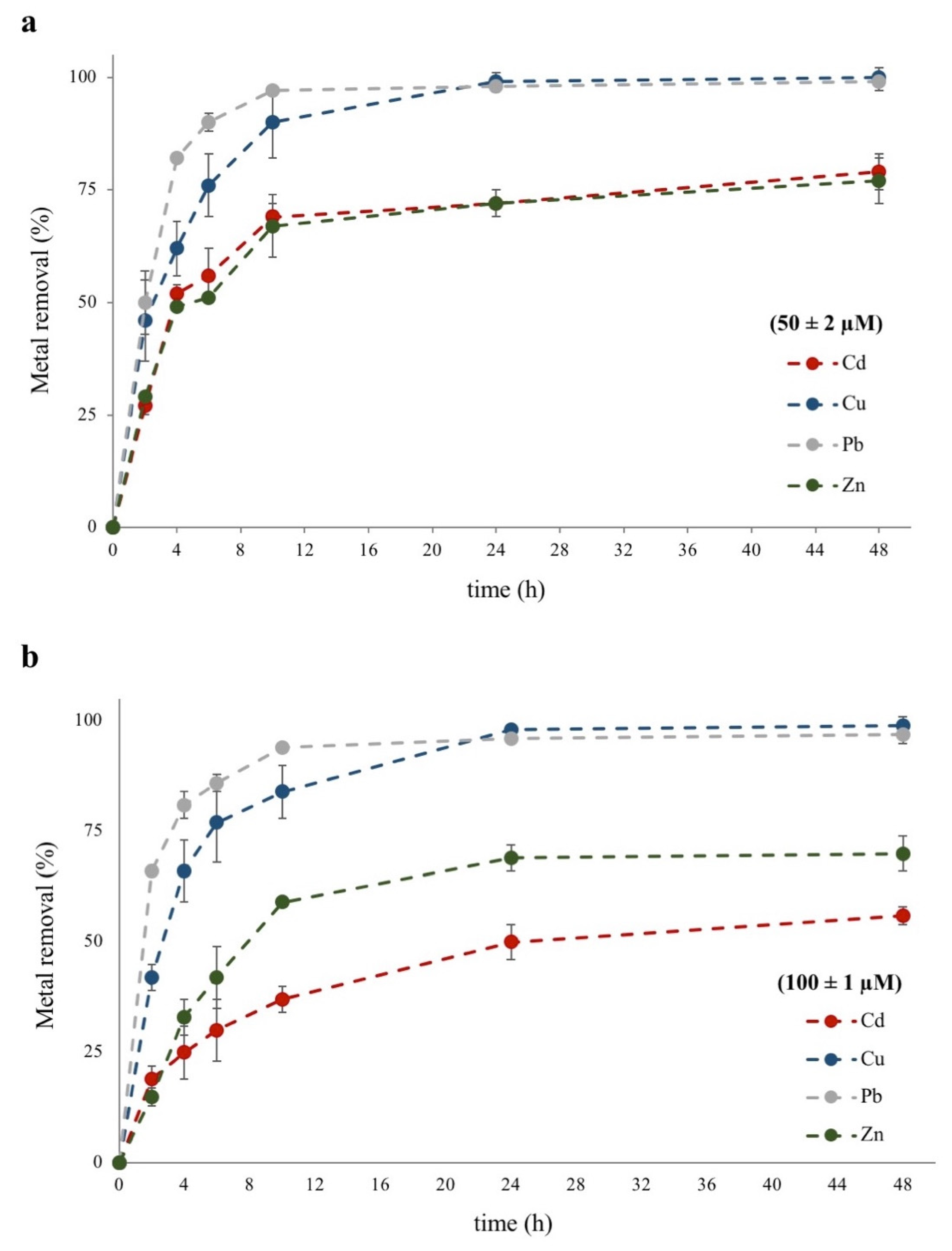
3.1. Metal Removal Efficiency from Water-Polluted Samples

Figure 1 shows the efficiency of naturally-occurring S. biloba specimens to remove Cd, Cu, Pb, and Zn from water artificially contaminated with 50 ± 2 µM (Figure 1a) or 100 ± 1 µM (Figure 1b) of metal, in independent assays during 48 h of plants exposure. A biphasic nature of metal elimination from the water column can be appraised with a first rapid phase process up to a period of about 6 h, and a slower one from 10 h onwards that reach a plateau between 24–48 h, depending on the metal and/or its concentration (Table 1). Similar patterns have been described by other authors for the removal of several metal ions using a wide variety of aquatic macrophytes, including S. biloba [12,14,15,18].
The non-linear kinetics of Cd, Cu, Pb, and Zn elimination from the water column observed in Figure 1, suggests that S. biloba utilizes various mechanisms for the removal of these cations. Metal sorption by the submerged root-like modified fronds (commonly named “roots”) of Salvinia specimens is probably the fastest component of metal uptake by these plants and may occur by a combination of physical and chemical processes such as ionic bonds, chemical chelation, and ionic exchange [15,18]. The content of macromolecules (i.e., carbohydrates, protein, and lipids) with negatively charged groups (e.g., carboxyl, sulphate, and phosphate) at the plant’s surface area, are key factors that determine the binding mechanism between the cationic metals and the biomass [25]. In such sense, Salvinia sp. biomass is characterized by a great specific surface area rich in carbohydrates and carboxyl groups, which may explain its high metal removal capacity [26,27]. Recently, [18] obtained the FTIR-ATR (Fourier transform infrared spectroscopy-Attenuated total reflectance) spectrum for S. biloba biomass and reported the presence of different functional groups such as carboxyl, phosphate, amide, and hydroxyl suggesting the involvement of these groups in Pb adsorption.
On the other hand, biological processes that involve metal intracellular uptake by transmembrane proteins or ionic channels and the posterior translocation to the aerial parts of the plant are likely responsible for the slower phase of metal removal in S. biloba [12,15,18].
Additionally, the data depicted in Figure 1a,b show that metal removal efficiency in S. biloba is also affected by both metal concentration and exposure time, in agreement with previous reports [12,13,14,15,16,17,18,19]. However, the phytoremediation efficiencies of S. biloba for Cd and Zn were strongly affected by the metal concentration in comparison to the performance observed for Cu and Pb.
As displayed in Table 1, in the first 6 h of plants exposure to 50 ± 2 µM of Cu or Pb, the metal removal efficiency was markedly high for both metals (76 ± 7% and 90 ± 2% for Cu and Pb, respectively). Noticeably, when Cu and Pb concentrations were doubled to 100 ± 1 µM, the efficiency of the bioprocess during the first 6 h was not practically affected since the removal rates of both metals were similar to those attained at the lower concentration (i.e., 50 ± 2 µM). Moreover, S. biloba showed a great capacity to remove Cu and Pb at both molar concentrations tested (>95%) in just 24 h of metal exposure, in agreement with the Cu and Pb hyperaccumulation capacity reported for these macrophytes [17,18]. However, as depicted in Figure 1, Cu elimination from the water column seemed to be slightly slower than that of Pb. This behavior is supported by the results shown in Table 1, where higher Pb removal values by S. biloba were reached between 4 to 10 h of metal-exposure, in comparison to the removal rates obtained for Cu at both 50 ± 2 µM and 100 ± 1 µM at the same period. Moreover, it should be highlighted that maximal Pb removal rates by S. biloba were obtained from 10 h onwards of metal exposure at both concentrations tested. These results agree with the lower rate constant K1 values calculated for Cu (Table 2, Section 3.2), suggesting some differences in Cu and Pb uptake mechanisms by this plant. In this context, [28] reported the activation of mechanisms of chelation and metal sequestration mediated by phytochelatins in the roots of S. minima exposed during 24 h to 40 µM Pb. More recently, [29] demonstrated an increment in the expression levels of four genes coding transmembrane transport proteins (SmABCC, SmATPase, SmNhaD, and SmABCG) in S. minima also exposed to 40 µM Pb during 24 h. Also, these authors have suggested that the above-mentioned genes are implicated in Pb accumulation in S. minima [29]. Hence, it may be expected that other Salvinia species share similar biosorption mechanisms for Pb uptake.
Regarding copper, the occurrence of metallothioneins (i.e., Cu-metallothioneins) and their importance for its detoxification have been reported in several organisms including plants, bacteria, yeast, mollusks, and mammals [30]. Moreover, [31] demonstrated a mild expression of a type 2 metallothionein gene (AzMT2) in the aquatic fern Azolla filiculoides exposed during 48 h to a final concentration of 10 µM, 100 µM, and 1 mM Cu, suggesting a role for AzMT2 in metal homeostasis. However, the presence of metallothioneins in Salvinia sp. has not yet been described.
Noticeably, S. biloba was less efficient removing Cd and Zn from polluted-water when compared to the performance observed for Cu and Pb. As exhibited in Table 1, after 6 h of plants exposure to 50 ± 2 µM Cd or Zn, metal removal efficiencies were 56 ± 6% and 51 ± 1%, respectively. These values were much lower than those obtained at the same time and conditions for Cu and Pb (76 ± 7% and 90 ± 2%, respectively). Furthermore, at 24–48 h metal exposition, S. biloba was able to remove less than 80% of both metals in contrast to its efficiency with 50 ± 2 µM Cu or Pb (>95%) (Table 1).
When Cd and Zn concentrations were doubled to 100 ± 1 µM, S. biloba showed significantly lower removal rates compared to those obtained at the lower concentration for both ions (Table 1). Moreover, Cd phytoremediation was the most affected by the metal concentration increase. As presented in Table 1, after 48 h metal exposition, S. biloba Zn removal efficiency decreased only 10% after doubling metal concentration in the sample (77 ± 5% for 50 ± 2 µM vs. 70 ± 4% for 100 ± 1 µM), whereas this decrease was approximately 40% when Cd concentration was doubled (from 79 ± 4% for 50 ± 2 µM to 56 ± 2% for 100 ± 1 µM). These results may be partly explained by the high Cd toxicity reported for several macrophytes, including Salvinia sp., even if at lower concentrations than those used at the present study [32,33,34].
Accordingly, all these data strongly suggest that S. biloba has lower efficiency to remove equal molar concentrations of Cd and Zn in comparison to Cu and Pb. However, it is interesting to note that at both tested concentrations, remnant Pb, Cu, and Zn in water samples were below the limit of admissible metal discharge in industrial wastewater (<2.4 µM for Pb, <23 µM for Cu, and <76 µM for Zn) established by resolution No. 1089/82 (Santa Fe, Argentina).
These variations in the capacity of S. biloba to phytoremediate Cd, Cu, Pb, or Zn from polluted-water should be explained by differences in the physical-chemical constants of the numerous physico-chemical equilibria (e.g., adsorption, chelation, ion exchange) involved at the early stages of metal uptake by the plants’ roots. In addition, different biological-regulated processes such as the expression of metalloproteins may also occur. Finally, metal-selective metabolic and/or physiological responses in S. biloba could also exist, and such responses may not only be dependent on the metal but also on its concentration, and to a lesser extent, on the exposure time to the contaminant.

3.2. Metal Removal Kinetics

Kinetics studies are significant for wastewater treatment as these allow predicted rates of contaminant removal to guide systems design. Also, the plants’ physiological need for metals plays a key role in the uptake kinetics of metal and directly or indirectly affects the metal accumulation process. Numerous kinetic models have been used to describe the reaction order of metal elimination in biological-regulated systems [21,35]. In most cases, the dynamics of such bioprocesses can be properly described by the first- or second-order reaction models [18,20].
As shown in Table 2, Cu and Pb removal from water fit a first-order reaction model, indicating that the rate of Cu and Pb phytoremediation by S. biloba depends mainly on metal initial concentration. In addition, the calculated K1 values for Pb removal were higher than the corresponding values obtained for Cu suggesting that Pb elimination by S. biloba is faster than Cu removal. These results confirm our previous observations (see Section 3.1).
Conversely, Cd and Zn removal from water fit a second-order reaction model, suggesting that the rate of the phytoremediation process may depend on two first-order reactants such as metal concentration and the number of metal-binding sites at S. biloba biomass.
Additionally, the rate constants K1 and K2 were always higher at the lowest concentration tested, indicating that metal concentration negatively affects the phytoremediation rate (i.e., metal removal by S. biloba becomes slower as the metal concentration in water increased). However, the removal rate of Cu and Pb by S. biloba was slightly affected when metal concentration was twice as great, while K2 values for Cd and Zn showed a 4- to 7-fold decrease.

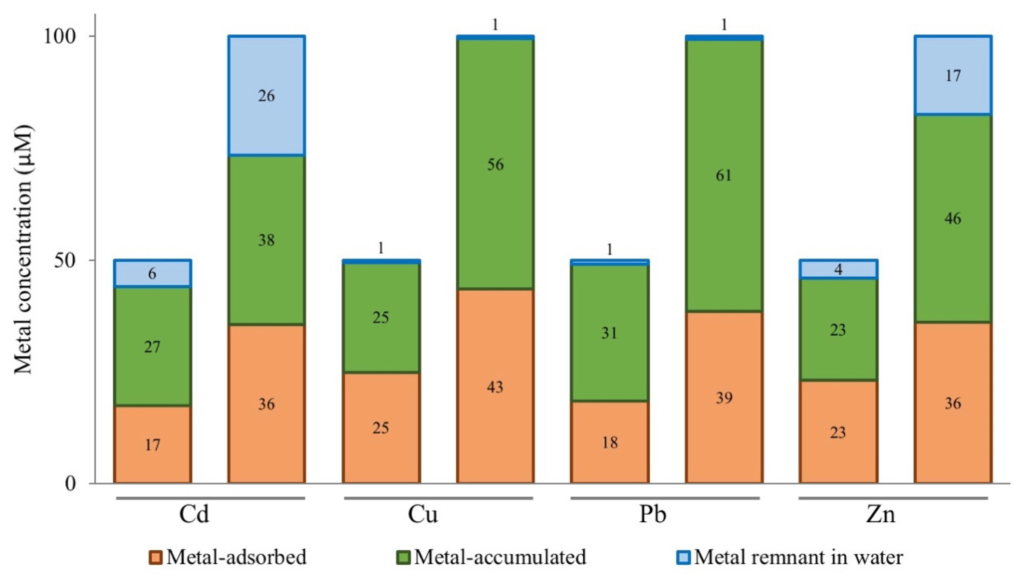
3.3. Elemental Analysis

To understand the fate of the metals within S. biloba microcosm after 48 h exposition to Cu, Pb, Cd, or Zn, three compartments were defined as described in [18]: (i) Remainder in the water column, (ii) adsorbed on the plant biomass surface, and (iii) accumulated into total plant biomass. As laid out in Figure 2, the greatest amount of metal removed from the water column was accumulated onto plant biomass, indicating that metal uptake within the plant cells was the main removal mechanism used by S. biloba under the assayed conditions. In general, the amount of metal accumulated was higher as the initial concentration of metal in the water sample increased. However, the intracellular molar concentrations of Cu, Pb, Cd, or Zn were very different according to each ion.
As expected, high intracellular values of Cu and Pb were found in S. biloba biomass in line with its high removal efficiencies (Figure 1 and Table 1). After 48 h of 100 ± 1 µM metal exposure, 2050 μg Cu/g and 4800 μg Pb/g were found in plant biomass according with the Cu and Pb hyperaccumulation capacity reported for S. biloba [17,18].
It is worth noting that, considering the lower efficiency showed for Cd and Zn removal by S. biloba (Table 1), the intracellular molar concentration for 50 ± 2 µM Cd or Zn treatment were not much different from the values found for Cu and Pb at the same treatment concentration (Figure 2). However, when heavy metal concentration was twice as high in the water samples, the differences in the amount of metal accumulated at S. biloba biomass were more evident. Moreover, a greater difference in the metal-adsorbed/metal-accumulated ratio was observed between Cd and Zn. As seen in Figure 2, the molar amount of Cd adsorbed on S. biloba biomass doubled when Cd concentration was two-fold, while its intracellular molar concentration showed a 40% increase. Contrarily, the opposite was virtually observed when increasing Zn concentration. In such case, doubling Zn concentration in water from 50 ± 2 µM to 100 ± 2 µM also doubled the amount of metal accumulated on plant biomass, while Zn-adsorbed showed a 55% increase. Besides, it should be noticed that the bioaccumulation capacity of Zn by S. biloba would not be fully appraised by only taking into account the biomass related to the amount of metal accumulated since at the highest concentration tested (i.e., 100 ± 2 µM) only 1300 µg Zn/g biomass could be calculated compared to 2100 µg Cd/g. These results show that weighing the competence of a plant species for metal-phytoremediation in terms of the ions accumulated in its biomass is not always representative of the total amount of ions removed since for those elements with low atomic weight this capacity can be undervalued.
As abovementioned (Section 3.1), the content of carbohydrates, protein, and lipids with carboxyl, sulphate, phosphate, thiol, and hydroxyl groups at S. biloba surface are key factors that determine the binding mechanism between the cationic metals and the plant’s biomass [18,25]. Thus, the presence of multiple metal-binding sites, and/or with different affinities, for Cu, Pb, Cd, and Zn onto S. biloba biomass is likely to occur. Moreover, metals penetrate plants mainly through the root system where most of these ions are bound to the cell wall. Therefore, the polysaccharides cellulose, lignin, and hemicellulose play an essential role in the complexation of metals [36,37]. Metal sequestration by cysteine- and glutamic acid-rich peptides such as glutathione, phytochelatins, and metallothioneins are some of the best well-known mechanisms of metal detoxification in plants. These peptides bind toxic heavy metals forming complexes that are stored into cell compartments such as vacuoles and chloroplasts, thus reducing the deleterious effect of metal ions to cells [31,38]. It has been proposed that in vivo, phytochelatins and metallothioneins are involved in the cellular detoxification and accumulation of several metals, including Cu, Pb, Cd, Ni, and Zn due to their ability to form stable metal-protein complexes [31,39,40,41]. In addition, [28] reported the induction of phytochelatins expression in roots and, to a lower extent, in leaves of S. minima in response to Pb; while [27] have suggested that calcium channels may also be involved in the intracellular accumulation of some divalent metals in S. minima. However, more experimental evidence is needed to support this hypothesis.
In conclusion, metal phytoremediation in S. biloba should be explained by a combination of several coordinated mechanisms that involve: (i) metal union (i.e., adsorption) to plant biomass, (ii) metal translocation through the cell wall, and (iii) metal sequestration, transport, and storage (e.g., into vacuoles and chloroplast). Therefore, the differences observed in the adsorbed-to-accumulated ratio for Cu, Pb, Cd, and Zn in S. biloba may depend on the type and number of metal-binding sites and their affinity constants, the stoichiometry of such interactions, the induction of metalloproteins (for chelation, transport, or sequestration), the opening of ionic channels (with possible differences in the migration rate for each ion), and even with steric impediments associated with the size of the cations. Although surface metal adsorption and intracellular accumulation are the main mechanisms used by S. biloba to phytoremediate metal-polluted water, more studies are still needed, at a molecular level, to elucidate the real complexity of such mechanisms.

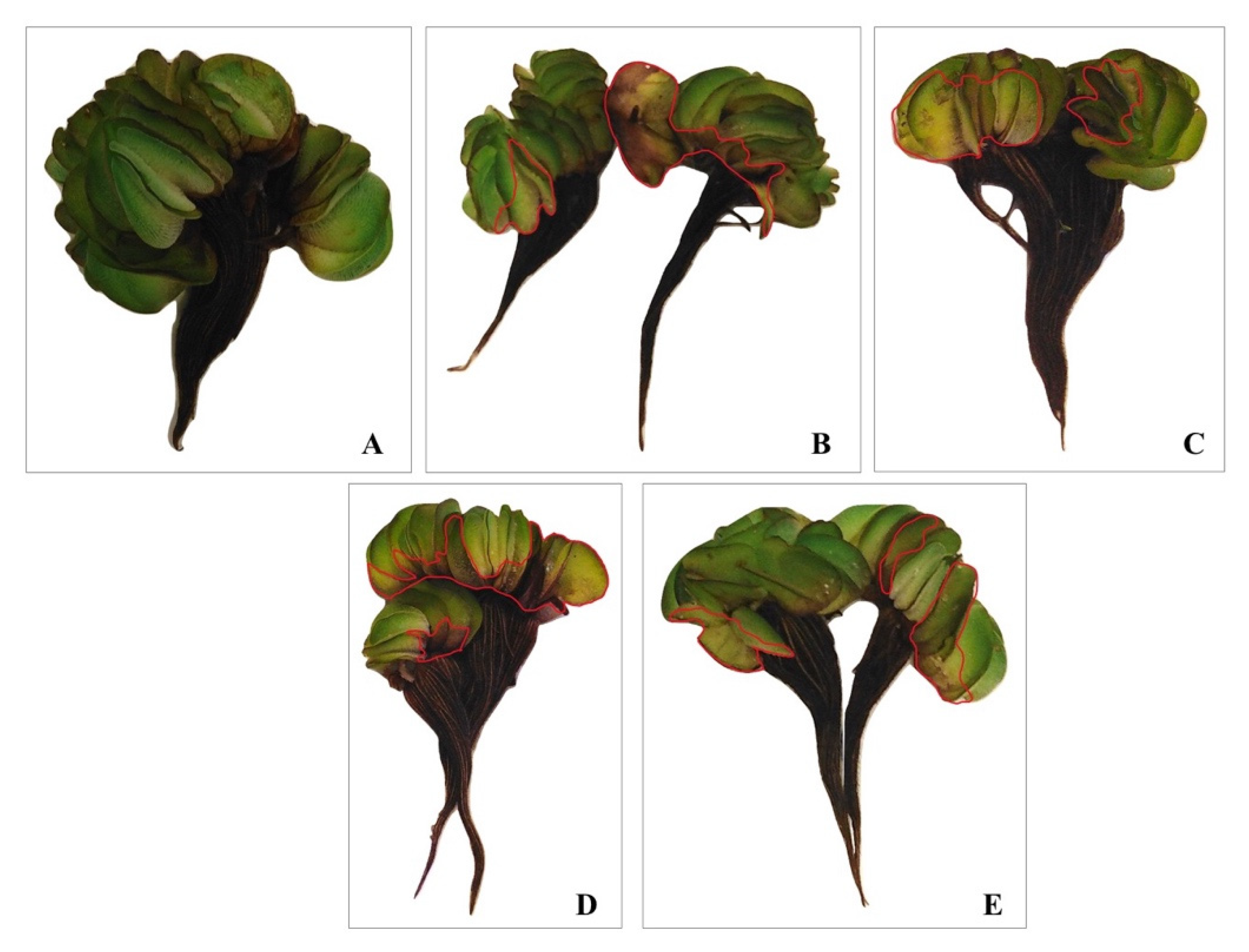
3.4. Evaluation of Metal Phytotoxicity in S. Biloba

S. biloba exposed to Cu, Pb, Cd, or Zn during 48 h presented some symptoms of metal phytotoxicity such as the presence of yellowish areas in leaves (i.e., chlorosis), alterations in the size and shape of the floating-fronds, and signs of necrosis. The occurrence of necrotized areas (i.e., dark brown pigmentation) on the surface of leaves increased with an increasing metal concentration in water samples (Figure 3). All these observations are in tune with the suggested use of Salvinia sp. as an ecological indicator of metal presence in contaminated environments [17,18,19,33].
Table 3 summarizes the data obtained for the content of chlorophylls, carotenoids, flavonoids, anthocyanins, and soluble carbohydrate in floating leaves of S. biloba specimens after 48 h exposure to 50 ± 2 µM or 100 ± 1 µM of Cu, Pb, Cd or Zn, to quantify metal-phytotoxicity. Noticeably, the four metals differently affected the distinct physiological markers evaluated. Chlorophylls content (chl a, chl b, and chl total) was significantly reduced (p < 0.05) after 48 h exposure to 50 ± 2 µM and 100 ± 1 µM of Cu or Pb. However, no differences were observed in the chlorophylls content for Cd and Zn treated-plants in comparison to the control specimens (i.e., no metal-exposed). These results suggest that both Cu and Pb interfere with the normal metabolism of S. biloba since chlorophylls play an important role in the capture of sunlight for plants photosynthetic bioprocesses [42]. In addition, carotenoids levels were also significantly affected when these macrophytes were exposed to 100 ± 1 µM Cu. Carotenoids are also essential to photosynthesis, acting as secondary photosynthetic pigments [43]. By contrast, carotenoids content increased in leaves of S. biloba after 48 h exposure to both 50 ± 2 µM and 100 ± 1 µM Cd. These results could explain in part the preservation in chlorophylls levels observed in leaves of S. biloba after Cd exposure since carotenoids can quench excess excitation energy protecting chlorophyll molecules from the oxidative damage [44,45]. No significant changes were observed for chlorophylls and carotenoids content in leaves of S. biloba exposed to Zn at both concentrations tested (Table 3). Although Zn-mediated alterations of the photosynthetic performance in other Salvinia species have been described [46], plants tolerance to Zn is an inheritable physiological property since this element is an essential micronutrient [47,48]. Moreover, plants physiological needs for Zn may be different even between closely related species.
Inversely, the concentration of flavonoids and anthocyanins in S. biloba leaves after 48 h exposure to water contaminated with 50 ± 2 µM and 100 ± 1 µM of Cu, Pb, Cd or Zn does not evidence any significant differences (Table 3). Phenolic compounds can act as metal ions chelating agents preventing the generation of radical oxygen species induced by the action of different metals [49]. Moreover, some related studies have reported an increase in the total amount of flavonoids and anthocyanins in Salvinia sp. exposed to metal(loids) indicating that these compounds could be involved in metal tolerance described for these aquatic ferns [19,23,50]. However, in our study, no increase in antioxidant pigments content was observed, possibly because the amount of Cu, Cd, Pb, and Zn accumulated into S. biloba leaves was not detrimental at the oxidative level.
Similar results were observed for the content of soluble carbohydrates (Table 3). It has been described that the presence of heavy metals in the environment affects the production of soluble carbohydrates in some species of Salvinia, resulting in a decrease of the macrophytes photosynthetic capacity [42,51]. However, our results indicate that the presence of Cu, Pb, Cd, or Zn at the selected concentrations and exposure times, did not affect carbohydrate metabolism in the aerial organs of S. biloba.
Altogether, these results suggest that S. biloba features different responses to counteract Cu, Cd, Pb, and Zn phytotoxicity. Although elements like Cu and Zn are critical for normal plant growth and development, high concentrations of essential as well as non-essential metals (e.g., Cd and Pb), can result in growth inhibition, chlorosis, water imbalance, among other toxicity symptoms. Plants possess a range of potential cellular mechanisms that may be involved in the detoxification of heavy metals and thus tolerance to metal stress. Some of such mechanisms include metal binding to the cell wall and extracellular exudates, reduced metal-uptake or efflux pumping of metals; peptide-mediated metal chelation (e.g., glutathione, phytochelatins, metallothioneins), and compartmentation of metals in the vacuole, among others [52]. Therefore, the tolerance of a plant against heavy metal stress is controlled by a complex and highly interrelated network of different molecular and physiological approaches that help to counteract metal phytotoxicity [53]. Some of such mechanisms are strictly directed to restrict metal translocation and transport to the most photosynthetically active organs of the plants (i.e., leaves), thus protecting them from the oxidative damage and harmful effects caused by the accumulation of heavy metals [19,52].

4. Conclusions

In this study, naturally-occurring S. biloba macrophytes obtained from Middle Paraná River (Entre Ríos, Argentina) were analyzed to evaluate their efficiency to phytoremediate water artificially contaminated with equal molar concentrations (50 ± 2 and 100 ± 1 µM) of Cd, Cu, Pb or Zn. S. biloba proved to be highly effective for Cu ad Pb removal (>95%) from water samples, and to a lesser extend for Zn (77–70%) and Cd (79–54%) elimination. The accumulation of Cd, Cu, Pb, and Zn in plant tissue induced evident visual alterations in leaves, supporting the use of this aquatic fern as an ecological indicator of heavy metal presence in contaminated waters. Additionally, some of the selected physiological parameters at S. biloba leaves were more sensitive than others to the harmful effects of metal accumulation. In general, the photosynthetic pigments were more susceptible to Cu and Pb stress, whereas carotenoids were increased by Cd exposure and decreased when plants were treated with 100 ± 1 µM Cu during 48 h. In addition, no changes in the content of antioxidant pigments (flavonoids and anthocyanins) and soluble carbohydrates in S. biloba leaves were observed. Therefore, the presented results demonstrate that S. biloba can differently manage equal levels of Cd, Cu, Pb, and Zn in water and display dissimilar physiological responses to counteract metal phytotoxicity. However, large-scale performance assays and long-term metal phytoremediation studies using S. biloba are still needed to further design plant-based systems for industrial-scale wastewater management.

Author Contributions

Conceptualization, L.M.S. and L.M.P.; methodology, W.G.L.O., J.E. and C.D.B.; formal analysis, J.E. and L.M.P.; investigation, W.G.L.O., J.E. and C.D.B.; writing—original draft preparation, L.A.B.N. and L.M.P.; writing—review and editing, L.A.B.N. and L.M.P.; supervision, L.M.P.; project administration, L.M.S. and L.M.P.; funding acquisition, L.M.S. and L.M.P. All authors have read and agreed to the published version of the manuscript.

Funding

This research was funded by the Pontifical Catholic University of Argentina (UCA-campus Rosario), grant number PROAPI-UCA 2019-2022; and the Portuguese Foundation for Science and Technology (FCT), grant number UIDB/04035/2020.

Acknowledgments

W.G.L.O. wishes to thank the National Scholarship and Educational Credit Program (PRONABEC) of the Ministry of Education of Peru for the funds provided. J.E. and C.D.B. thank the Vicerrectorado de Investigación e Innovación Académica (Pontifical Catholic University of Argentina-UCA) for the financial support.

Conflicts of Interest

The authors declare no conflict of interest. The funders had no role in the design of the study; in the collection, analyses, or interpretation of data; in the writing of the manuscript, or in the decision to publish the results.

References

  1. Jaishankar, M.; Tseten, T.; Anbalagan, N.; Mathew, B.B.; Beeregowda, K.N. Toxicity, mechanism and health effects of some heavy metals. Interdiscip. Toxicol. 2014, 7, 60–72. [Google Scholar] [CrossRef] [PubMed] [Green Version]
  2. Azimi, A.; Azari, A.; Rezakazemi, M.; Ansarpour, M. Removal of heavy metals from industrial wastewaters: A review. ChemBioEng Rev. 2017, 4, 37–59. [Google Scholar] [CrossRef]
  3. Ali, H.; Khan, E.; Sajad, M.A. Phytoremediation of heavy metals—Concepts and applications. Chemosphere 2013, 91, 869–881. [Google Scholar] [CrossRef] [PubMed]
  4. Barakat, M.A. New trends in removing heavy metals from industrial wastewater. Arab. J. Chem. 2011, 4, 361–377. [Google Scholar] [CrossRef] [Green Version]
  5. Sukumaran, D. Phytoremediation of heavy metals from industrial effluent using constructed wetland technology. Appl. Ecol. Environ. Sci. 2013, 1, 92–97. [Google Scholar] [CrossRef]
  6. Sharma, P.; Pandey, S. Status of phytoremediation in world scenario. Int. J. Environ. Biorem. Biodeg. 2014, 2, 178–191. [Google Scholar] [CrossRef]
  7. Dhir, B. Potential of biological materials for removing heavy metals from wastewater. Environ. Sci. Pollut. Res. Int. 2014, 21, 1614–1627. [Google Scholar] [CrossRef]
  8. Dixit, R.; Wasiullah, E.; Malaviya, D.; Pandiyan, K.; Singh, U.B.; Sahu, A.; Shukla, R.; Singh, B.P.; Rai, J.P.; Sharma, P.K.; et al. Bioremediation of heavy metals from soil and aquatic environment: An overview of principles and criteria of fundamental processes. Sustainability 2015, 7, 2189–2212. [Google Scholar] [CrossRef] [Green Version]
  9. Rai, P.K. Heavy metal phytoremediation from aquatic ecosystems with special reference to macrophytes. Crit. Rev. Environ. Sci. Technol. 2009, 39, 697–753. [Google Scholar] [CrossRef]
  10. Marchetti, Z.Y.; Latrubesse, E.M.; Pereira, M.S.; Ramonell, C.G. Vegetation and its relationship with geomorphologic units in the Parana River floodplain, Argentina. J. S. Am. Earth Sci. 2013, 46, 122–136. [Google Scholar] [CrossRef]
  11. Dhir, B. Salvinia: An aquatic fern with potential use in phytoremediation. Environ. We Int. J. Sci. Technol. 2009, 4, 23–27. [Google Scholar]
  12. Maine, M.A.; Duarte, M.; Suñe, N.L. Cadmium uptake by floating macrophytes. Water Res. 2001, 35, 2629–2634. [Google Scholar] [CrossRef]
  13. Maine, M.A.; Suñe, N.L.; Lagger, S.C. Chromium bioaccumulation: Comparison of the capacity of two floating aquatic macrophytes. Water Res. 2004, 38, 1494–1501. [Google Scholar] [CrossRef] [PubMed]
  14. Paris, C.; Hadad, H.R.; Maine, M.A.; Suñe, A.N. Eficiencia de dos macrófitas flotantes libres en la absorción de metales pesados. Limnetica 2005, 24, 237–244. [Google Scholar] [CrossRef]
  15. Suñe, N.L.; Sánchez Galván, G.; Caffaratti, S.; Maine, M.A. Cadmium and chromium removal kinetics from solution by two aquatic macrophytes. Environ. Pollut. 2007, 145, 467–473. [Google Scholar] [CrossRef]
  16. Hadad, H.R.; Maine, M.A.; Natale, G.S.; Bonetto, C. The effect of nutrient addition on metal tolerance in Salvinia herzogii. Ecol. Eng. 2007, 31, 122–131. [Google Scholar] [CrossRef]
  17. Freitas, F.; Lunardi, S.; Souza, L.B.; von der Osten, J.S.C.; Arruda, R.; Andrade, R.L.T.; Battirola, L.D. Accumulation of copper by the aquatic macrophyte Salvinia biloba Raddi (Salviniaceae). Braz. J. Biol. 2018, 78, 133–139. [Google Scholar] [CrossRef] [Green Version]
  18. Tello Zevallos, W.; Salvatierra, L.M.; Loureiro, D.B.; Morató, J.; Pérez, L.M. Evaluation of the autochthonous free-floating macrophyte Salvinia biloba Raddi for use in the phytoremediation of water contaminated. Desalin. Water Treat. 2018, 103, 282–289. [Google Scholar] [CrossRef] [Green Version]
  19. Castillo Loría, K.; Emiliani, J.; Herrero, M.S.; Bergara, C.D.; Salvatierra, L.M.; Pérez, L.M. Effect of daily exposure to Pb-contaminated water into Salvinia biloba physiology and phytoremediation performance. Aquatic Toxicol. 2019, 210, 158–166. [Google Scholar] [CrossRef]
  20. Liu, Z.-R.; Chen, X.-S.; Zhou, L.-M.; Wei, P. Development of a first-order kinetics-based model for the adsorption of nickel onto peat. Min. Sci. Technol. 2009, 19, 230–234. [Google Scholar] [CrossRef]
  21. Zheng, X.; Bi, C.; Li, Z.; Podariu, M.; Hage, D.S. Analytical methods for kinetic studies of biological interactions: A review. J. Pharm. Biomed. Anal. 2015, 113, 163–180. [Google Scholar] [CrossRef] [Green Version]
  22. Wintermans, J.F.; De Mots, A. Spectrophotometric characteristics of chlorophyll a and b and their phenophytins in ethanol. Biochim. Biophys. Acta 1965, 109, 448–455. [Google Scholar] [CrossRef]
  23. Bizzo, A.L.; Intorne, A.C.; Gomes, P.H.; Susuki, M.S.; Esteves, B.D. Short-term physiological responses to copper stress in Salvinia auriculata Aubl. Acta Limnol. Bras. 2014, 26, 268–277. [Google Scholar] [CrossRef] [Green Version]
  24. Casati, P.; Walbot, V. Differential accumulation of maysin and rhamnosylisoorientin in leaves of high-altitude landraces of maize after UV-B exposure. Plant Cell Environ. 2005, 28, 788–799. [Google Scholar] [CrossRef]
  25. Kratochvil, D.; Volesky, B. Advances in the biosorption of heavy metals. Trends Biotechnol. 1998, 16, 291–300. [Google Scholar] [CrossRef]
  26. Sánchez-Galván, G.; Monroy, O.; Gómez, J.; Olguín, E.J. Assessment of the hyperaccumulating lead capacity of Salvinia minima using bioadsorption and intracellular accumulation factors. Water Air Soil Pollut. 2008, 194, 77–90. [Google Scholar] [CrossRef]
  27. Olguín, E.J.; Sánchez-Galván, G.; Pérez-Pérez, T.; Pérez-Orozco, A. Surface adsorption, intracellular accumulation and compartmentalization of lead in batch-operated lagoons with Salvinia minima as affected by environmental conditions, EDTA and nutrients. J. Ind. Microbiol. Biotechnol. 2005, 32, 577–586. [Google Scholar] [CrossRef] [PubMed]
  28. Estrella-Gómez, N.; Mendoza-Cózatl, D.; Romero-Sánchez, R.; Gonzáles-Mendoza, D.; Zapata-Pérez, O.; Martínez-Hernández, A.; Santamaría, J.M. The Pb-hyperaccumulator aquatic fern Salvinia minima Baker, responds to Pb2+ by increasing phytochelatins via changes in SmPCS expression and in phytochelatin synthase activity. Aquatic Toxicol. 2009, 91, 320–328. [Google Scholar] [CrossRef]
  29. Leal-Alvarado, D.A.; Estrella-Maldonado, H.; Sáenz-Carbonell, L.; Ramírez-Prado, J.H.; Zapata-Pérez, O.; Santamaría, J.M. Genes coding for transporters showed a rapid and sharp increase in their expression in response to lead, in the aquatic fern (Salvinia minima Baker). Ecotoxicol. Environ. Saf. 2018, 147, 1056–1064. [Google Scholar] [CrossRef]
  30. Calvo, J.; Jung, H.; Meloni, G. Copper metallothioneins. IUBMB Life 2017, 69, 236–245. [Google Scholar] [CrossRef] [Green Version]
  31. Schor-Fumbarov, T.; Goldsbrough, P.; Adam, Z.; Tel-Or, E. Characterization and expression of a metallothionein gene in the aquatic fern Azolla filiculoides under heavy metal stress. Planta 2005, 223, 69–76. [Google Scholar] [CrossRef]
  32. Phetsombat, S.; Kruatrachue, M.; Pokethitiyook, P.; Upatham, S. Toxicity and bioaccumulation of cadmium and lead in Salvinia cucullata. J. Environ. Biol. 2006, 27, 645–652. [Google Scholar] [PubMed]
  33. Wolff, G.; Pereira, G.C.; Castro, E.M.; Louzada, J.; Coelho, F.F. The use of Salvinia auriculata as a bioindicator in aquatic ecosystems: Biomass and structure dependent on the cadmium concentration. Braz. J. Biol. 2012, 72, 71–77. [Google Scholar] [CrossRef] [PubMed] [Green Version]
  34. Rodrigues, D.A.; Vasconcelos Filho, S.C.; Rodrigues, A.A.; Rampazzo, D.F.; Rodrigues, C.L.; Vasconcelos, J.M.; Magalhães, P.A.N.R. Effect of cadmium on the morphology and anatomy of Salvinia auriculata. Afr. J. Biotechnol. 2016, 15, 891–896. [Google Scholar] [CrossRef] [Green Version]
  35. Rsat, H.; Petzold, L.; Pettigrew, M.F. Kinetic modeling of biological systems. Methods Mol. Biol. 2009, 541, 311–335. [Google Scholar] [CrossRef] [Green Version]
  36. Joseph, L.; Jun, B.M.; Flora, J.R.V.; Park, C.M.; Yoon, Y. Removal of heavy metals from water sources in the developing world using low-cost materials: A review. Chemosphere 2019, 229, 142–159. [Google Scholar] [CrossRef] [PubMed]
  37. Yu, H.; Wang, J.; Yu, J.; Wang, Y.; Chi, R. Effects of surface modification on heavy metal adsorption performance and stability of peanut shell and its extracts of cellulose, lignin, and hemicellulose. Environ. Sci. Pollut. Res. 2020. [Google Scholar] [CrossRef] [PubMed]
  38. Cobbett, C.S. Phytochelatins and their roles in heavy metal detoxification. Plant Physiol. 2000, 123, 825–832. [Google Scholar] [CrossRef] [Green Version]
  39. Mendoza-Cózatl, D.; Rodríguez-Zavala, J.; Rodríguez-Enríquez, S.; Mendoza-Hernández, G.; Briones-Gallardo, G.; Moreno-Sánchez, R. Phytochelatin-cadmium-sulfide high-molecular-mass complexes of Euglena gracilis. FEBS J. 2006, 273, 5703–5713. [Google Scholar] [CrossRef]
  40. Mendoza-Cózatl, D.; Rangel-González, E.; Moreno-Sánchez, R. Simultaneous Cd2+, Zn2+, and Pb2+ uptake and accumulation by photosynthetic Euglena gracilis. Arch. Environ. Contam. Toxicol. 2006, 51, 521–528. [Google Scholar] [CrossRef]
  41. Clemens, S. Toxic metal accumulation, responses to exposure and mechanisms of tolerance in plants. Biochimie 2006, 88, 1707–1719. [Google Scholar] [CrossRef]
  42. Leal-Alvarado, D.A.; Espadas, F.; Sáenz-Carbonell, L.; Talavera, C. Lead accumulation reduces photosynthesis in the lead hyper-accumulator Salvinia minima Baker by affecting the cell membrane and inducing stomatal closure. Aquatic Toxicol. 2016, 171, 37–47. [Google Scholar] [CrossRef] [PubMed]
  43. Gomes, S.M.D.S.; De Lima, V.L.A.; De Souza, A.P.; Do Nascimento, J.J.V.R.; Do Nascimento, E.S. Chloroplast pigments as indicators of lead stress. Eng. Agríc. 2014, 34, 877–884. [Google Scholar] [CrossRef] [Green Version]
  44. Ramel, F.; Mialoundama, A.S.; Havaux, M. Nomenzymic carotenoid oxidation and photooxidative stress signalling in plants. J. Exp. Bot. 2013, 64, 799–805. [Google Scholar] [CrossRef] [PubMed]
  45. Munné-Bosch, S.; Queval, G.; Foyer, C.H. The impact of global change factors on redox signaling underpinning stress tolerance. Plant Physiol. 2013, 161, 5–19. [Google Scholar] [CrossRef] [Green Version]
  46. Dhir, B.; Sharmila, P.; Pardha-Saradhi, P. Photosynthetic performance of Salvinia natans exposed to chromium and zinc rich wastewater. Braz. J. Plant Physiol. 2008, 20, 61–70. [Google Scholar] [CrossRef]
  47. Chaney, R.L. Zinc Phytotoxicity. In Zinc in Soils and Plants; Robson, A.D., Ed.; Springer: Dordrecht, The Netherlands, 1993; Volume 55, pp. 135–150. [Google Scholar] [CrossRef]
  48. Broadley, M.R.; White, P.J.; Hammond, J.P.; Zelko, I.; Lux, A. Zinc in plants. New Phytol. 2007, 173, 677–702. [Google Scholar] [CrossRef]
  49. Prado, C.; Ponce, S.C.; Pagano, E.; Prado, F.E.; Rosa, M. Differential physiological responses of two Salvinia species to hexavalent chromium at a glance. Aquatic Toxicol. 2016, 175, 213–221. [Google Scholar] [CrossRef]
  50. Michalak, A. Phenolic compounds and their antioxidant activity in plants growing under heavy metal stress. Polish J. Environ. Stud. 2006, 15, 523–530. [Google Scholar]
  51. Dhir, B.; Sharmila, P.; Pardha-Saradhi, P.; Sharma, P.; Kumar, R.; Mehta, D. Heavy metal induced physiological alterations in Salvinia natans. Ecotoxicol. Environ. Saf. 2011, 74, 1678–1684. [Google Scholar] [CrossRef]
  52. Hall, J.L. Cellular mechanisms for heavy metal detoxification and tolerance. J. Exp. Bot. 2002, 53, 1–11. [Google Scholar] [CrossRef] [PubMed]
  53. Singh, S.; Parihar, P.; Singh, R.; Singh, V.P.; Prasad, S.M. Heavy metal tolerance in plants: Role of transcriptomics, proteomics, metabolomics, and ionomics. Front. Plant Sci. 2016, 6, 1143. [Google Scholar] [CrossRef] [PubMed] [Green Version]
Figure 1. Metal removal efficiency by Salvinia biloba specimens exposed during 48 h to water contaminated with Cu, Cd, Pb, or Zn at (a) 50 ± 2 µM or (b) 100 ± 1 µM. Data are mean ± SD of three independent experiments (n = 3).
Water 12 01737 g001
Figure 2. Metal distribution (µM) in S. biloba microcosms after 48 h exposure to metal-polluted water with 50 ± 2 µM or 100 ± 1 µM of Cu, Pb, Cd, or Zn (n = 3).
Water 12 01737 g002
Figure 3. Phenotypic changes observed in S. biloba specimens after 48 h exposure to metal-polluted water: control (A); 100 ± 1 µM Cd (B), Cu (C), Pb (D), or Zn (E). Red-marked areas indicate some representative changes on leaves (chlorosis and signs of necrosis) described in the text.
Water 12 01737 g003
Table 1. Percentage of removed Cd, Cu, Pb, and Zn by S. biloba at different exposure times and metal initial concentrations.
Exposure Time (h) 50 ± 2 µM100 ± 1 µM
CdCuPbZnCdCuPbZn
00A0A0A0A0A0A0A0A
227 ± 2bB46 ± 9cB50 ± 7cB29 ± 1bB19 ± 3aB42 ± 3cB66 ± 1dB15 ± 2aB
452 ± 2bC62 ± 6cC82 ± 1dC49 ± 1bC25 ± 6aBC66 ± 7cBC81 ± 3dC33 ± 4aC
656 ± 6bC76 ± 7cCD90 ± 2dD51 ± 1bC30 ± 7aC77 ± 9cCD86 ± 2cdD42 ± 7abC
1069 ± 3cD 90 ± 8deDE97 ± 1eE67 ± 7cD37 ± 3aC84 ± 6dD94 ± 1eE59 ± 1bD
2472 ± 1bDE99 ± 2cE98 ± 1cE72 ± 3bDE50 ± 4aD98 ± 1cE96 ± 1cE69 ± 3bDE
4879 ± 4cE100 ± 2dE99 ± 2dE77 ± 5cE56 ± 2aD99 ± 2dE97 ± 2dE70 ± 4bE
1 Results are presented as mean ± S.D. Different lowercase letters in the same row indicate significant statistical difference (p < 0.05) between different metals and concentrations; e.g., “a” and “b” are statistically different from each other but not from “ab”. Different uppercase letters in the same file indicate significant statistical difference (p < 0.05) between exposure time for each metal at each concentration; e.g., “A” and “B” are statistically different from each other but not from “AB”.
Table 2. Rate constants of first (K1) and second (K2) order kinetic models obtained for Cu, Pb, Cd, and Zn removal from the metal-polluted water column by S. biloba.
Metal (µM)First-order KineticsSecond-order Kinetics
K1 (1/h)R2K2 (L/mg.h)R2
Cd50 ± 20.11630.92270.05770.9922
100 ± 10.04240.89210.00800.9932
Cu50 ± 20.18590.99361.34340.8932
100 ± 10.17700.99050.60330.8842
Pb50 ± 20.39860.99070.19910.9340
100 ± 10.31730.99500.09450.8399
Zn50 ± 20.10200.94130.09390.9985
100 ± 10.09280.93620.02300.9946
Table 3. Content of photosynthetic (chlorophylls and carotenoids) and antioxidants (flavonoids y anthocyanins) pigments, and soluble carbohydrates in leaves of S. biloba after 48 h exposure to metal-polluted water with 50 ± 2 µM or 100 ± 1 of Cd, Pb, Cu, or Zn.
Physiological ParametersControlCd (µM)Cu (µM)Pb (µM)Zn (µM)
50 ± 2 100 ± 1 50 ± 2100 ± 1 50 ± 2 100 ± 1 50 ± 2 100 ± 1
chl a
(µg/g FW)
156 ± 7a
(100%)
151 ± 15a
(97%)
160 ± 19a
(102%)
138 ± 24ab
(88%)
102 ± 16b
(65%)
123 ± 10b
(79%)
113 ± 5b
(72%)
156 ± 18a
(100%)
166 ± 36a
(106%)
chl b
(µg/g FW)
109 ± 6a
(100%)
92 ± 8a
(84%)
92 ±7a
(84%)
78 ± 12b
(71%)
65 ± 9b
(62%)
82 ± 7b
(75%)
81 ± 9b
(74%)
109 ± 14a
(100%)
105 ± 22a
(101%)
chl total
(µg/g FW)
261 ± 14a
(100%)
242 ± 24a
(93%)
257 ± 11a
(98%)
198 ± 35b
(76%)
167 ± 25b
(64%)
205 ± 16b
(78%)
194 ± 14b
(74%)
265 ± 25a
(102%)
271 ± 41a
(104%)
Carotenoids
(µg/g FW)
35 ± 2a
(100%)
43 ± 3b
(123%)
43 ± 2b
(123%)
37 ± 2a
(106%)
28 ± 2b
(80%)
36 ± 3a
(103%)
37 ± 2a
(106%)
34 ± 2a
(97%)
38 ± 4a
(108%)
Flavonoids
(Abs/g FW)
87 ± 5a
(100%)
89 ± 4a
(102%)
82 ±8a
(94%)
91 ± 8a
(105%)
84 ± 4a
(97%)
95 ± 8a
(109%)
86 ± 7a
(99%)
93 ± 7a
(107%)
83 ± 7a
(95%)
Anthocyanins
(Abs/g FW)
560 ± 46a
(100%)
584 ± 45a
(104%)
549 ± 75a
(98%)
545 ± 8a
(97%)
596 ± 37a
(106%)
603 ± 78a
(108%)
566 ± 45a
(101%)
610 ± 60a
(109%)
581 ± 50a
(104%)
Carbohydrates
(mg/g FW)
42 ± 1a
(100%)
43 ± 3a
(102%)
41 ± 5a
(98%)
40 ± 7a
(95%)
38 ± 9a
(90%)
46 ± 2a
(109%)
37 ± 2a
(88%)
41 ± 7a
(98%)
40 ± 7a
(95%)
1 Results are presented as mean ± S.D. FW—Fresh Weight. Different letters in the same row indicate significant statistical difference (p < 0.05); e.g., “a” and “b” are statistically different from each other but not from “ab”. Numbers in brackets are the percent mean values calculated with respect to the control group.

