Until recently, the study of human anatomy has looked at muscle function as isolated units responsible for specific movements. While a muscle can perform a discrete function when used in a particular exercise, generally speaking the only time that a muscle works in isolation is when using a machine designed to focus on a specific body part with a predetermined range of motion. Most movements required for activities of daily living (ADL), popular sports or recreational activities feature patterns that involve numerous muscles working together to create the desired actions.
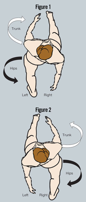
Popular exercises for the adductor muscles include lying on the side to lift the leg off the floor or sitting in a machine that requires squeezing the legs together. While these exercises might create the perception that the adductors are being targeted, these movements are actually not using the adductors the way they are designed to work as humans move during the gait cycle. When the right femur is flexed during the gait cycle (with the leg in front of the body; Figure 1), and the left femur is extended (with the leg in back of the body), gravity causes a frontal plane shift in the pelvis, creating adduction in the right hip and abduction in the left. As the right femur (and leg) transitions to extension and the left leg swings forward, the pelvis shifts to the left to create abduction in the right femur and adduction in the left (Figure 2). This happens with every step, so adduction does occur, but only as a result of the frontal plane motion of the pelvis.
The purpose of this ongoing functional anatomy series is to help health and fitness professionals develop an understanding of how muscles actually function as a component of an integrated system. This second installment focuses on the adductors of the inner thigh, an area of the body that many clients are interested in targeting. However, many of the exercises commonly used to “tone” this area don’t use these muscles in the way they are actually designed to function on our bodies.
Understanding How Muscles Function
Traditional anatomy often teaches that certain muscles work on only one joint or to create a specific movement. However, when it comes to performing integrated motor patterns, many muscles work together to produce efficient and coordinated movements. Gait, either walking or running, is the foundational pattern of human movement and is an excellent example of how muscles work synergistically to create coordinated movements. In the human body, muscles, connective tissue and skeletal structures are aligned in such a way to make gait as efficient as possible.
Understanding how muscles function to create the human gait cycle can help you design and deliver exercise programs that address the way that muscles actually work, thus delivering the results that clients and class participants seek. No other muscle group is more misunderstood than the adductor complex. Too often, people use exercises designed to isolate the adductor muscle group of the inner thigh with movements that focus on femoral adduction, or bringing the thighs closer to the midline of the body. But take a step back and think of how humans walk: Do we consciously bring our thighs closer to the midline of the body every time we take a step? No, we don’t. The femurs do adduct toward the midline of the body, but only as the result of how the pelvis moves in the frontal plane as the body shifts its weight from one leg to the other.
The Adductor Complex
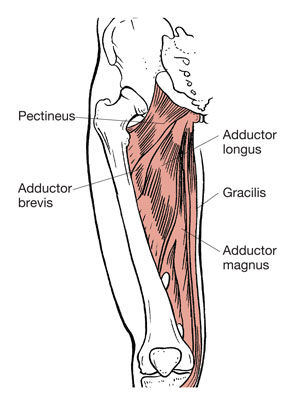
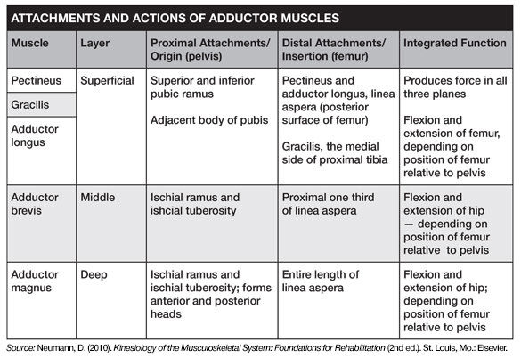
The adductor muscle group can be organized into three distinct layers. The superficial layer includes the pectineus, adductor longus and gracilis. The adductor brevis comprises the middle layer, while the deep layer consists of the large, triangular adductor magnus (Neumann, 2010). It can be challenging to see how the muscles attach to the femur when viewing two-dimensional images in a textbook. The adductors do not attach to the medial border of the bone; rather, they have insertions along the linea aspera, which is along the posterior aspect of the femur. As a result, when the femur is in an extended position the adductors are lengthened and create flexion of the iliofemoral (hip) joint when shortened. Likewise, when the hip is flexed the adductors are lengthened to create extension of the iliofemoral joint as the muscles contract. When in a neutral, anatomical position, the adductors can pull the femurs closer to the midline of the body, but this motion may not help them to become more efficient in controlling flexion and extension of the hip.
Understanding Movement of the Adductor Complex
Understanding how muscles work to produce movement during gait can provide essential insights into how muscles are designed to function. This is especially important in the case of the adductors. The full gait cycle is comprised of one complete movement for each leg. A single phase of the cycle starts when the right foot makes contact with the ground (the heel-strike phase) and the left foot is preparing to leave the ground (the heel-off phase). When the body is passing over the right foot (the mid-stance phase), the left foot is swinging forward (the leg-swing phase). Once each leg has transitioned from heel strike to mid-stance to heel-off, one full cycle is complete. The adductors are involved with the entire cycle. When the leg is forward in heel-strike, the adductors function to pull the femur into hip extension; when the leg is extended in heel-off, the adductors pull the femur forward into hip flexion. The specific length of time of each phase of gait is as follows (American Council on Exercise, 2015):
- Double-leg support, when both feet are on the ground
- Single-leg support, when balanced on the right leg as the left swings forward
- Double-leg support, when both feet are again in contact with the ground
- Single-leg support, when the body is balanced on the left leg as the right leg swings forward
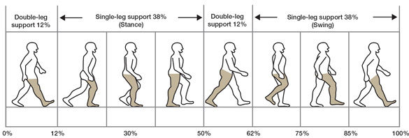
The only time the adductors are not actively involved in flexing or extending the femur is during the moment the body is directly over the foot during the mid-stance phase. The adductors, specifically the magnus and longus, are the most active muscle group during the gait cycle, with both the adductor magnus and longus active during the 76 percent of the time when the body is balanced on one leg. It’s important to note that between 40 and 70 degrees of hip flexion, the line of force runs through the medial-lateral axis of the body (the axis of rotation for movement in the sagittal plane), where the adductor muscles lose the ability to generate torque in the sagittal plane. Outside of this 30-degree arc, the adductors provide significant torque as both flexors and extensors (Neumann, 2010).
Muscle is comprised of two types of tissue:
- The contractile element of the actin and myosin proteins, which, according to the sliding filament theory, overlap to create the shortening force for muscle contractions
- The non-contractile component of the fascia and elastic connective tissue, which surround every individual muscle fiber.
As a muscle is lengthened, it stores elastic potential energy, which is then released as mechanical energy when the muscle shortens. Understanding the role of fascia and elastic connective tissue in storing and releasing energy is important for knowing how the adductors work during the gait cycle (Schleip and Baker, 2015; Earls, 2014).
The adductor magnus is most active during heel contact (as the front foot is making contact with the ground) and during mid-stance (when the body is balanced on a single leg while the other leg is swinging). As the body is balanced on the left leg, the left adductor magnus is helping pull the leg into extension as the right-side adductor magnus is pulling the right hip into flexion and then decelerating flexion as the lower leg extends to prepare to make contact with the ground (Earls, 2014; Neumann, 2010).
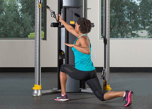
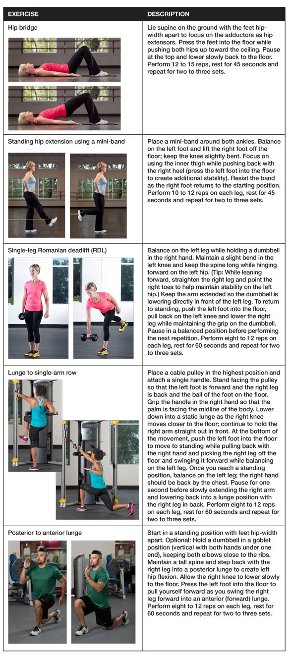
Exercises to Strengthen the Adductor Complex
The adductors work to create movement of the femur under the pelvis during the leg swing phase, while also controlling motion of the pelvis over a relatively fixed leg during the single-leg support phase. When it comes to strength-training exercises for the adductors, the key concept is to focus on moving in the sagittal plane in a closed-chain position with the feet firmly planted on the ground. The adductors are lengthened when the hip is both extended and flexed; therefore, it is important to strengthen the muscles during both the lengthening and shortening phases of muscle action.
Summary
Understanding how the adductors function to move the legs through both flexion and extension during the gait cycle helps to explain why many athletes who play soccer, lacrosse, rugby or field hockey have such well-defined legs. It is possible that the rapid acceleration and deceleration that occurs when athletes are constantly reacting to the movements of both teammates and opponents places tremendous forces on the adductors, which results in hypertrophy and definition. Understanding how the adductors function during gait will enable you to develop a variety of exercises to help create the strong, lean and toned legs many clients wish to achieve.
References
American Council on Exercise (2015). ACE Medical Exercise Specialist Manual, San Diego, Calif.: American Council on Exercise.
Earls, J. (2014). Born to Walk: Myofascial Efficiency and the Body in Movement. Berkeley, Calif.: Lotus Publishing.
Neumann, D. (2010). Kinesiology of the Musculoskeletal System: Foundations for Rehabilitation (2nd ed.). St. Louis, Mo.: Elsevier.
Schleip, R. and Baker, A. (2015). Fascia in Sport and Movement. Edinburgh, Scotland: Handspring Publishing.
 by
by