Thrombolytic therapy
Show Alternative Names
Tissue plasminogen activator
TPA
Alteplase
Reteplase
Tenecteplase
Activase thrombolytic agent
Clot-dissolving agents
Reperfusion therapy
Stroke - thrombolytic
Heart attack - thrombolytic
Acute embolism - thrombolytic
Thrombosis - thrombolytic
Lanoteplase
Staphylokinase
Streptokinase (SK)
Urokinase
Stroke - thrombolytic therapy
Heart attack - thrombolytic therapy
Stroke - thrombolysis
Heart attack - thrombolysis
Myocardial infarction - thrombolysis
Thrombolytic therapy is the use of drugs to break up or dissolve blood clots, which are the main cause of both heart attacks and stroke.
Information
Thrombolytic medicines are approved for the emergency treatment of stroke and heart attack. The most commonly used drug for thrombolytic therapy is tissue plasminogen activator (tPA), but other drugs can do the same thing.
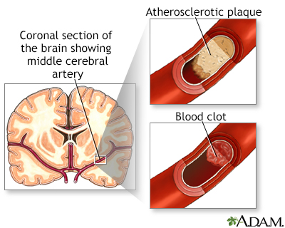
Stroke
A stroke occurs when blood flow to a part of the brain stops. A stroke is sometimes called a "brain attack. " If blood flow is cut off for longer th...

Image
Read Article Now
Book Mark Article
Heart attack
Most heart attacks are caused by a blood clot that blocks one of the coronary arteries. The coronary arteries bring blood and oxygen to the heart. ...

Image
Read Article Now
Book Mark Article
Ideally, you should receive thrombolytic medicines within the first 30 minutes after arriving at the hospital for treatment.
Cardiac and vascular disorders: Blood clots - Animation
A thrombus is a blood clot that forms inside a blood vessel, often causing a blockage of the flow of blood through the vascular system. The process of thrombus formation, called “thrombosis”, results from the activation of the normal blood clotting system. In pathological thrombosis, the process of coagulation far exceeds the body’s capacity to dissolve the clot. As additional layers of platelets and fibrin are deposited, the thrombus continues to grow larger until a mass is formed. This thrombus can eventually block the space within a blood vessel called the “lumen”, resulting in complete blockage. An embolus is the sudden blockage of an artery by a mass that is carried through the bloodstream, eventually lodging in a blood vessel that is too small to allow its passage. The mass can be composed of fat, cancer cells, air, or even a blood clot. For example, a thromboembolism is a thrombus that detached and traveled through the circulatory system eventually blocking a distant artery. When a long bone is fractured, or during orthopedic surgery, damaged fat cells in the bone marrow may release fragments of fat into the bloodstream – that can form a fat embolism. Similarly, large fragments of bone marrow including hematopoietic tissue may be released into the bloodstream, which can cause a bone marrow embolism. Clumps of cancer cells may break free into the circulation to form tumor emboli. An air embolus can form as a result of the inadvertent introduction of air bubbles during a catheterization procedure. There are two main types of embolus depending on whether it lodges in the pulmonary or systemic circulation. A pulmonary embolus originates in peripheral veins and is carried to the heart where it enters the pulmonary circulation, eventually becoming lodged in the pulmonary arterial system. A systemic embolus originates in the heart and travels via the aorta to the systemic arterial circulation. They commonly affect the arteries leading to the brain, kidneys, spleen, gut, and lower limbs.
HEART ATTACKS
A blood clot can block the arteries to the heart. This can cause a heart attack, when part of the heart muscle dies due to a lack of oxygen being delivered by the blood.
Thrombolytics work by dissolving a major clot quickly. This helps restart blood flow to the heart and helps prevent damage to the heart muscle. Thrombolytics can stop a heart attack that would otherwise be larger or potentially deadly. Outcomes are better if you receive a thrombolytic drug within 12 hours after the heart attack starts. But the sooner treatment begins, the better the results.
The drug restores some blood flow to the heart in most people. However, the blood flow may not be completely normal and there may still be a small amount of muscle damaged. Further therapy, such as cardiac catheterization with angioplasty and stenting, may be needed.
Your health care provider will base the decisions about whether to give you a thrombolytic medicine for a heart attack on many factors. These factors include your history of chest pain and the results of an ECG test.
ECG test
An electrocardiogram (ECG) is a test that records the electrical activity of the heart.

Image
Read Article Now
Book Mark Article
Other factors used to determine if you are a good candidate for thrombolytics include:
- Age (older people are at increased risk of complications)
- Sex
- Medical history (including your history of a previous heart attack, diabetes, low blood pressure, or increased heart rate)
Generally, thrombolytics may not be given if you have:
- A recent head injury
- Bleeding problems
- Bleeding ulcers
- Pregnancy
- Recent surgery
- Taken blood thinning medicines such as Coumadin
- Trauma
- Uncontrolled (severe) high blood pressure
STROKES
Most strokes are caused when blood clots move to a blood vessel in the brain and block blood flow to that area. For such strokes (ischemic strokes), thrombolytics can be used to help dissolve the clot quickly. Giving thrombolytics within 3 hours of the first stroke symptoms can help limit stroke damage and disability.
The decision to give the drug is based upon:
- A brain CT scan to make sure there has not been any bleeding
- A physical exam that shows a significant stroke
- Your medical history
As in heart attacks, a clot-dissolving drug isn't usually given if you have one of the other medical problems listed above.
Thrombolytics are not given to someone who is having a stroke that involves bleeding in the brain. They could worsen the stroke by causing increased bleeding.
OTHER USES
Thrombolytic medications are sometimes used to treat other conditions including:
- Acute pulmonary embolism
Acute pulmonary embolism
A pulmonary embolus is a blockage of an artery in the lungs. The most common cause of the blockage is a blood clot.
 ImageRead Article Now Book Mark Article
 - Clots in vascular catheters such as those used for dialysis
RISKS
Bleeding is the most common risk. It can be life threatening.
Minor bleeding from the gums or nose can occur in approximately 25% of people who receive the drug. Bleeding into the brain occurs approximately 1% of the time. This risk is the same for both stroke and heart attack patients.
If thrombolytics are felt to be too dangerous, other possible treatments for clots causing a stroke or heart attack include:
- Removal of the clot (thrombectomy)
- A procedure to open narrowed or blocked blood vessels that supply blood to the heart or the brain
CONTACT A HEALTH CARE PROVIDER OR CALL 911
Heart attacks and strokes are medical emergencies. The sooner treatment with thrombolytics begins, the better the chance for a good outcome.
Review Date:
5/8/2022
Reviewed By
Michael A. Chen, MD, PhD, Associate Professor of Medicine, Division of Cardiology, Harborview Medical Center, University of Washington Medical School, Seattle, WA. Also reviewed by David C. Dugdale, MD, Medical Director, Brenda Conaway, Editorial Director, and the A.D.A.M. Editorial team.
References
Bohula EA, Morrow DA. ST-elevation myocardial infarction: management. In: Libby P, Bonow RO, Mann DL, Tomaselli GF, Bhatt DL, Solomon SD, eds. Braunwald's Heart Disease: A Textbook of Cardiovascular Medicine. 12th ed. Philadelphia, PA: Elsevier; 2022:chap 38.
Jaffer IH, Weitz JI. Antithrombotic drugs. In: Hoffman R, Benz EJ, Silberstein LE, et al, eds. Hematology: Basic Principles and Practice. 7th ed. Philadelphia, PA: Elsevier; 2018:chap 149.
Kabrhel C. Pulmonary embolism and deep vein thrombosis. In: Walls RM, Hockberger RS, Gausche-Hill M, eds. Rosen's Emergency Medicine: Concepts and Clinical Practice. 10th ed. Philadelphia, PA: Elsevier; 2023:chap 74.
Kuo WT. Percutaneous interventions for acute pulmonary embolism. In: Mauro MA, Murphy KP, Thomson KR, Venbrux AC, Morgan RA, eds. Image-Guided Interventions. 3rd ed. Philadelphia, PA: Elsevier; 2021:chap 55.
O'Gara PT, Kushner FG, Ascheim DD, et al. 2013 ACCF/AHA guideline for the management of ST-elevation myocardial infarction: a report of the American College of Cardiology Foundation/American Heart Association Task Force on Practice Guidelines. Circulation. 2013;127(4):529-555. PMID: 23247303 pubmed.ncbi.nlm.nih.gov/23247303/.
Papa L, Meurer WJ. Stroke. In: Walls RM, Hockberger RS, Gausche-Hill M, eds. Rosen's Emergency Medicine: Concepts and Clinical Practice. 10th ed. Philadelphia, PA: Elsevier; 2023:chap 87.
Disclaimer
The information provided herein should not be used during any medical emergency or for the diagnosis or treatment of any medical condition. A licensed medical professional should be consulted for diagnosis and treatment of any and all medical conditions. Links to other sites are provided for information only -- they do not constitute endorsements of those other sites.
No warranty of any kind, either expressed or implied, is made as to the accuracy, reliability, timeliness, or correctness of any translations made by a third-party service of the information provided herein into any other language. © 1997-
A.D.A.M., a business unit of Ebix, Inc. Any duplication or distribution of the information contained herein is strictly prohibited.


 All rights reserved.