Prevalence of Arcobacter and Other Pathogenic Bacteria in River Water in Nepal
by
 , , ,
, , ,
Rajani Ghaju Shrestha
1,
Sarmila Tandukar
2 ,
,
Dinesh Bhandari
3,4,
Samendra P. Sherchan
5,
Yasuhiro Tanaka
6,
Jeevan B. Sherchand
3 and
Eiji Haramoto
1,*
1
Interdisciplinary Center for River Basin Environment, University of Yamanashi, 4-3-11 Takeda, Kofu, Yamanashi 400-8511, Japan
2
Department of Natural, Natural, Biotic and Social Environment Engineering, University of Yamanashi, 4-3-11 Takeda, Kofu, Yamanashi 400-8511, Japan
3
Institute of Medicine, Tribhuvan University, Maharajgunj, Kathmandu 1524, Nepal
4
School of Public Health, The University of Adelaide, Adelaide, South Australia 5005, Australia
5
Department of Global Environmental Health Sciences, Tulane University, 1440 Canal Street, Suite 2100, New Orleans, LA 70112, USA
6
Department of Environmental Sciences, University of Yamanashi, 4-4-37 Takeda, Kofu, Yamanashi 400-8510, Japan
*
Author to whom correspondence should be addressed.
Water 2019, 11(7), 1416; https://doi.org/10.3390/w11071416
Submission received: 11 June 2019
/
Revised: 5 July 2019
/
Accepted: 8 July 2019
/
Published: 10 July 2019
(This article belongs to the Section Water Quality and Contamination)
Abstract
:This study aims to determine the diversity of pathogenic bacteria in the Bagmati River, Nepal, during a one-year period. A total of 18 river water samples were collected from three sites (n = 6 per site) along the river. Bacterial DNA, which were extracted from the water samples, were analyzed for bacterial 16S rRNA genes by next-generation sequencing for 13 of 18 samples, and by quantitative PCR targeting Arcobacter for all 18 samples. The 16S rRNA sequencing identified an average of 97,412 ± 35,909 sequences/sample, which were then categorized into 28 phyla, 61 classes, and 709 bacterial genera. Eighteen (16%) genera of 111 potential pathogenic bacteria were detected with abundance ratios of >1%; Arcobacter, Acinetobacter, and Prevotella were the dominant genera. The Arcobacter abundance ratios were 28.6% (n = 1), 31.3 ± 15.8% (n = 6), and 31.8 ± 17.2% (n = 6) at the upstream, midstream, and downstream sites, respectively. Arcobacter was detected in 14 (78%) of 18 samples tested, with concentrations ranging from 6.7 to 10.7 log10 copies/100 mL, based on quantitative PCR. Our results demonstrate the poor bacterial quality of the Bagmati River water, suggesting a need for implementing more measures to reduce fecal contamination in the river water.
1. Introduction
Industrial pollution load, poor sewage and wastewater treatment facilities, inadequate water pollution control laws, and rapid urbanization have all contributed to the increased degradation of the river water environment in developing countries [1,2]. The Bagmati River, which flows from the northern boundary southwards across the urban areas of the Kathmandu Valley in Nepal, is suffering from chemical, biological, and solid waste pollution [1,3,4,5,6,7,8,9].
Arcobacter, which is widely recognized as an emerging waterborne and foodborne pathogen [10,11,12], has been detected in river water samples in Japan, Nepal, and Spain [4,13,14,15,16]. Arcobacter in river water samples has been linked to fecal contamination from animal or human sources [11,13]. In Nepal, only a few studies using next-generation sequencing (NGS) and quantitative PCR (qPCR) have detected Arcobacter in the river and groundwater samples [4,14]. The abundance ratios and concentrations of Arcobacter were highest in river water samples in these studies [4,14]. NGS has been a useful tool in obtaining large numbers of sequences to determine microbial diversity and community structure, as explained in a previous study that was conducted by Caporaso et al. (2012) [17].
A study that was conducted in three sites of the Bagmati River (Sundarijal (upstream), Thapathali (midstream), and Chovar (downstream)) reported that the microbiological quality of the river water upstream was less degraded when compared to those at the other two sites [9]. Indicator bacteria, potential index viruses, and human-fecal markers were more frequently detected than protozoa and human enteric viruses at all three Bagmati River sites. However, it is essential to also understand the bacterial community of river water at these sites. Therefore, this study aimed to determine the bacterial diversity and quantify Arcobacter at three Bagmati River sites.
2. Materials and Methods
2.1. Collection of River Water Samples and Extraction of Bacterial DNA
As previously described [9], the Bagmati River water samples were collected every two months from the upstream, midstream, and downstream regions during a one-year period between November 2015 and September 2016, as shown in Figure 1. The sampling sites were selected based on population density in three regions of the Bagmati River basin, where the population density is lower in the upstream region when compared to the midstream and downstream regions, and also to observe the difference in bacterial community in start and end point of the Bagmati River flowing through the urban area of the Kathmandu Valley. The water samples were collected in 100-mL sterile plastic bottles and transported to the laboratory in dry ice, kept at 4 °C, and then processed within 4 h.
Ten milliliters of the river water samples were filtered through a 0.22-µm filter paper, which was used to extract the bacterial DNA using CicaGeneus DNA Extraction Reagent (Kanto Chemical, Tokyo, Japan). Briefly, the membrane filter containing the microorganisms was resuspended in 5 mL of Tris-EDTA buffer (pH 7.4) into a 50 mL plastic tube. Bacterial DNA was extracted from the mixture using the CicaGeneus DNA extraction kit after shaking and vortex mixing at 50 °C with a speed of 300 rpm (Kanto Chemical). Bacterial DNA that were extracted from the river water samples were used both in a study that was conducted by Tandukar, et al., in 2018 as well as in this study [9].
2.2. NGS for the Characterization of Bacterial Communities
Of the 18 samples collected, 13 samples, excluding five of six samples from Sundarijal in which bacterial DNA concentrations were not enough high, were subjected to metagenomics sequencing using a MiSeq gene sequencer (Illumina, San Diego, CA, USA) as previously described [18]. Briefly, the 16S rRNA library was prepared, and the sequence data were analyzed after purification, amplification, library quantification, normalization, and pooling. Operational taxonomic units were analyzed at the bacterial domain, phylum, family, and genus levels. A genus was considered to be a potentially pathogenic bacterium if any one species of the genus was categorized as biosafety level 2 or 3 by the American Biological Safety Association (https://my.absa.org/tiki-index.php?page=Riskgroups), as previously described [4]. The raw sequences were registered in the National Center for Biotechnology Information (NCBI) Sequence Read Archive under the accession number PRJNA547952.
2.3. Quantification of Total Bacteria and Arcobacter
Total bacterial 16S rRNA genes were quantified by qPCR while using the 515F and U806R primers [19,20]. The qPCR mixture components were as follows: 2.0 µL of template DNA, 0.1 µL of 50 pmol/µL each of the forward and reverse primers, and 12.5 µL of SYBR Premix Ex TaqII (Tli RNaseH Plus) (Takara Bio, Otsu, Japan), with a final volume of 25.0 µL. The thermal cycling conditions were at 95 °C for 30 s, followed by 35 cycles at 94 °C for 15 s, 55 °C for 30 s, and 72 °C for 20 s, as previously described [4].
Arcobacter spp. were quantified by SYBR Green-based qPCR using a reaction volume of 25 µL containing 2.0 µL of template DNA, 12.5 µL of a MightyAmp for Real Time (SYBR Plus) (Takara Bio), 0.1 µL of 50 pmol/µL each of Arco-F and Arco-R-rev primers [14]. The thermal conditions were as follows: at 98 °C for 2 min, followed by 35 cycles at 98 °C for 10 s, at 55 °C for 30 s, and at 68 °C for 40 s, and finally at 95 °C for 15 s, at 60 °C for 30 s, and at 95 °C for 15 s to identify a specific single peak melting temperature (Tm) [14].
2.4. Statistical Analysis
The Pearson’s correlation coefficient test was performed with Microsoft Excel 2016 (Microsoft Corporation, Redmond, WA, USA) to determine the relationship of Arcobacter with fecal indicator bacteria, human-fecal markers, and index viruses [9]. Differences were considered to be statistically significant if the resulting p value was <0.05.
3. Results
3.1. Characterization of Bacterial Community by NGS
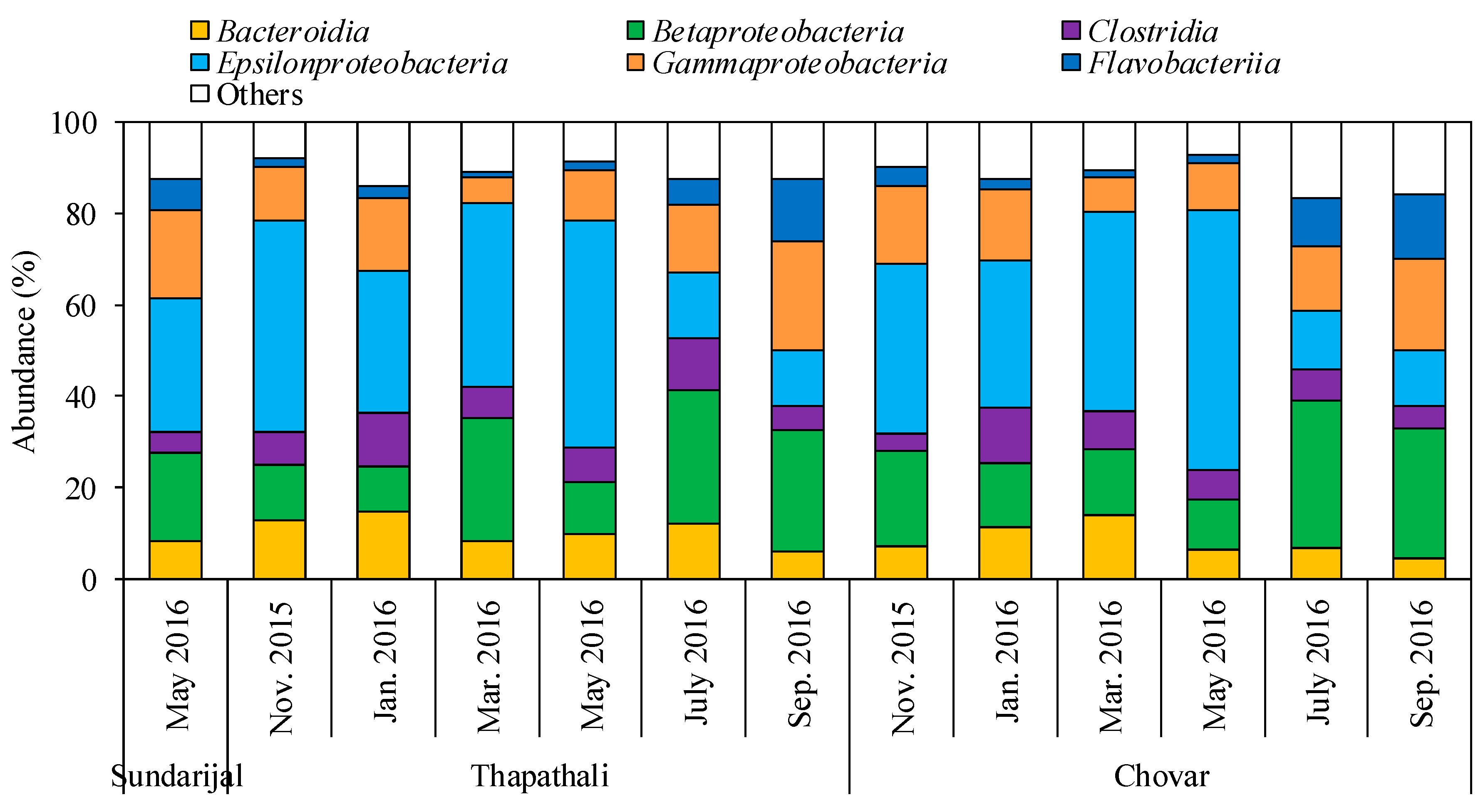
Thirteen water samples that were collected from three Bagmati River sites were subjected to the Illumina MiSeq sequencing, where the average number of 16S rRNA gene sequences obtained was 97,412 ± 35,909 per sample. As shown in Figure 2, among the 28 phyla identified, Proteobacteria (59–80%) was the most dominant in all of the samples, followed by Bacteroidetes (10–25%) and Firmicutes (6–15%). Epsilonproteobacteria (29–57%) was dominant in nine river water samples among the 61 classes that were detected, whereas Betaproteobacteria (27–32%) was dominant in four river water samples, as shown in Figure 3. The abundance ratios of classes Gammaproteobacteria and Bacteroidia in all of the samples tested ranged from 6–24% and 5–15%, respectively. Of the 111 potential pathogenic bacteria identified, 18 genera were detected at abundance ratios of >1% in at least one of the 13 samples tested, as summarized in Table 1. Arcobacter was the most dominant genus at all three sites, with abundance ratios of 28.6% (n = 1), 31.3 ± 15.8% (n = 6), and 31.8 ± 17.2% (n = 6) at Sundarijal, Thapathali, and Chovar, respectively. At Sundarijal, Thapathali, and Chovar, the abundance ratios of Acinetobacter were 10.3%, 5.3 ± 5.3%, and 5.0 ± 3.9%, respectively, whereas Prevotella was detected at abundance ratios of 5.9%, 6.8 ± 2.4%, and 4.6 ± 1.5%, respectively.
3.2. Occurrence of Total Bacteria and Arcobacter in River Water Samples
As shown in Figure 4, the total bacteria and Arcobacter levels in six river water samples each from Sundarijal, Thapathali, and Chovar were quantified using qPCR. The minimum and maximum concentrations of total bacteria at the three sites were 7.1 and 10.5 log10 copies/100 mL, respectively. In Sundarijal, Thapathali, and Chovar, Arcobacter was detected in two (33%), six (100%), and six (100%) river water samples, with concentrations of 6.7–10.4, 9.0–10.4, and 8.4–10.7 log10 copies/100 mL, respectively.
4. Discussion
Diverse groups of potential pathogenic bacteria, such as Arcobacter, Acinetobacter, and Prevotella, were detected by NGS in all three regions of the Bagmati River during a one-year sampling period. In this study, the abundance ratio of the class Epsilonproteobacteria was higher than that of class Betaproteobacteria, whereas Gammaproteobacteria had the highest abundance ratio in previous studies [4,14]. This may be attributed to differences in the time and place of sample collection. Although the bacterial community in the upstream Sundarijal sample was identified, the results of sequencing of only one sample will not be enough to represent a one-year sampling period, unlike Thapathali and Chovar.
Among the potential pathogenic bacterial 16S rRNA genes detected by NGS, those of Arcobacter were found in the highest abundance ratio. Comparable results have been reported in previous studies, although a limited number of river water samples were tested [4,14]. During a year of water sampling, Arcobacter was detected in all sampling months at Thapathali and Chovar. In a previous study, the highest Arcobacter concentration was found in a river water sample (9.1 log10 copies/100 mL) among the 13 Arcobacter-positive samples that were collected from shallow dug wells, deep tube wells, and a river [14]. The detection of Arcobacter in wastewater samples, irrigation water, sludge, drinking water, surface water, groundwater, and seawater [10,12] shows the bacterium’s capability to survive and multiply in water with a broad range of qualities.
People living in the Kathmandu Valley are highly dependent on groundwater use [21]. However, the poor availability of the required volume of water in the valley for domestic purposes leads to the use of polluted river water for irrigation and for washing vegetables [7,22]. The indirect consumption of such polluted river water contaminated with different enteric viruses, protozoa, and bacteria may have adverse effects on human health. The polluted river water not only pollutes its surroundings, but it also contaminates any nearby groundwater. Chemical analysis has provided evidence for the interconnectivity between groundwater and surface water, a link that allows for the exchange of chemicals between each water source [23]. The same interconnectivity of waterborne pathogens may thus occur, which increases the risk of waterborne diseases in the Kathmandu Valley.
There is evidence that the physical, chemical, and biological deterioration of water of the Bagmati River shows an alarming situation of river environment, which affects the surrounding environment. The lowest abundance ratio of Arcobacter at Sundarijal indicates that the upstream region has been less affected by anthropogenic pollution when compared to the midstream and downstream regions. This result is supported by a previous study that found lower concentrations of protozoa and human enteric viruses at Sundarijal than at Thapathali and Chovar [9]. In addition to the microbiological water quality, solid waste levels and the chemical quality of river water in the downstream region become worse as the river travels through urban areas [24,25].
The relationships of Arcobacter with fecal indicator bacteria (Escherichia coli), human-fecal markers (BK and JC polyomaviruses, and human Bacteroidales), and plant viruses (tobacco mosaic virus and pepper mild mottle virus) were determined using data from Tandukar et al. (2018) [9]. Index viruses were also included in this analysis because they have been proposed as an indicator of the prevalence of human enteric viruses in irrigation water sources in the Kathmandu Valley [7]. The Pearson’s correlation coefficient (r) values in Table 2 show the significant correlation between Arcobacter and E. coli, BK polyomaviruses, pepper mild mottle virus, and human Bacteroidales, but there was no significant correlation between Arcobacter and JC polyomaviruses and tobacco mosaic virus. The lack of correlation with the second group of viruses may be due to the difference in the properties between viruses and bacteria. The presence of Arcobacter, along with other fecal indicator bacteria, human-fecal markers, index viruses, and human enteric viruses in the river water samples, should not be neglected. Thus, the river water’s microbial quality needs to be studied more, and strict rules and regulations should be imposed in order to improve waste disposal and management in facilities impacting the river.
5. Conclusions
In summary, of the 111 potential pathogenic bacteria that were identified by NGS, 18 (16%) had abundance ratios of >1%. Arcobacter, Acinetobacter, and Prevotella were frequently detected at the three sites of the Bagmati River. The bacterial communities in upstream, midstream, and downstream of the Bagmati River were identified with a wide range of bacteria. Using qPCR, Arcobacter was successfully detected in 14 (78%) of 18 river water samples, at concentrations of 6.7–10.7 log10 copies/100 mL. Arcobacter was detected in all of the samples that were collected from the midstream and downstream sites of the Bagmati River. The prevalence of Arcobacter, along with other presumptive bacterial pathogens in river water samples, shows poor water quality, which suggests a need to prevent further fecal contamination of the river water.
Author Contributions
R.G.S. processed the samples, analyzed the results, and prepared a draft of the manuscript. S.T. and D.B. conceived the design of the study and processed the samples, S.P.S. processed the samples, Y.T. checked the analyzed results and corrected the draft of the manuscript, J.B.S. conceived the design of the study. E.H. conceived the design of the study, checked the analyzed results, and corrected the draft of the manuscript.
Funding
This study was supported by Kurita Water and Environment Foundation, the Science and Technology Research Partnership for Sustainable Development (SATREPS) program of Japan International Cooperation Agency (JICA) and Japan Science and Technology Agency (JST), entitled “Hydro-microbiological Approach for Water Security in Kathmandu Valley, Nepal”, and the Japan Society for the Promotion of Science (JSPS) through Fund for the Promotion of Joint International Research (Fostering Joint International Research (B) (grant number JP18KK0297)).
Acknowledgments
The authors thank Bijay Man Shakya (University of Yamanashi, Japan) for his help in map preparation.
Conflicts of Interest
The authors declare no conflict of interest.
References
- Kannel, P.R.; Lee, S.; Lee, Y.-S.; Kanel, S.R.; Pelletier, G.J. Application of automated QUAL2Kw for water quality modeling and management in the Bagmati River, Nepal. Ecol. Model. 2007, 202, 503–517. [Google Scholar] [CrossRef]
- Karn, S.K.; Harada, H. Surface water pollution in three urban territories of Nepal, India, and Bangladesh. Environ. Manag. 2001, 28, 483–496. [Google Scholar]
- Bhatt, M.P.; McDowell, W.H.; Gardner, K.H.; Hartmann, J. Chemistry of the heavily urbanized Bagmati River system in Kathmandu Valley, Nepal: Export of organic matter, nutrients, major ions, silica, and metals. Environ. Earth Sci. 2014, 71, 911–922. [Google Scholar] [CrossRef]
- Ghaju Shrestha, R.; Tanaka, Y.; Malla, B.; Bhandari, D.; Tandukar, S.; Inoue, D.; Sei, K.; Sherchand, J.B.; Haramoto, E. Next-generation sequencing identification of pathogenic bacterial genes and their relationship with fecal indicator bacteria in different water sources in the Kathmandu Valley, Nepal. Sci. Total Environ. 2017, 601–602, 278–284. [Google Scholar] [CrossRef] [PubMed]
- Haramoto, E.; Yamada, K.; Nishida, K. Prevalence of protozoa, viruses, coliphages and indicator bacteria in groundwater and river water in the Kathmandu Valley, Nepal. Trans. R. Soc. Trop. Med. Hyg. 2011, 105, 711–716. [Google Scholar] [CrossRef] [PubMed]
- Kannel, P.R.; Lee, S.; Kanel, S.R.; Khan, S.P.; Lee, Y.-S. Spatial–temporal variation and comparative assessment of water qualities of urban river system: A case study of the river Bagmati (Nepal). Environ. Monit. Assess. 2007, 129, 433–459. [Google Scholar] [CrossRef] [PubMed]
- Shrestha, S.; Shrestha, S.; Shindo, J.; Sherchand, J.B.; Haramoto, E. Virological quality of irrigation water sources and pepper mild mottle virus and tobacco mosaic virus as index of pathogenic virus contamination level. Food Environ. Virol. 2018, 10, 107–120. [Google Scholar] [CrossRef] [PubMed]
- Shrestha, S.; Haramoto, E.; Shindo, J. Assessing the infection risk of enteropathogens from consumption of raw vegetables washed with contaminated water in Kathmandu Valley, Nepal. J. Appl. Microbiol. 2017, 123, 1321–1334. [Google Scholar] [CrossRef]
- Tandukar, S.; Sherchand, J.B.; Bhandari, D.; Sherchan, S.P.; Malla, B.; Ghaju Shrestha, R.; Haramoto, E. Presence of human enteric viruses, protozoa, and indicators of pathogens in the Bagmati River, Nepal. Pathogens 2018, 7, 38. [Google Scholar] [CrossRef]
- Banting, G.; Figueras Salvat, M.J. Arcobacter. In Global Water Pathogens Project; Rose, J.B., Jiménez-Cisneros, B., Eds.; Michigan State University: E. Lansing, MI, USA; UNESCO: Paris, France, 2017; pp. 3–25. Available online: http://www.waterpathogens.org/book/arcobacter (accessed on 9 July 2019).
- Collado, L.; Figueras, M.J. Taxonomy, epidemiology, and clinical relevance of the genus Arcobacter. Clin. Microbiol. Rev. 2011, 24, 174–192. [Google Scholar] [CrossRef] [PubMed]
- Hsu, T.-T.D.; Lee, J. Global distribution and prevalence of Arcobacter in food and water. Zoonoses Public Health 2015, 62, 579–589. [Google Scholar] [CrossRef] [PubMed]
- Collado, L.; Inza, I.; Guarro, J.; Figueras, M.J. Presence of Arcobacter spp. in environmental waters correlates with high levels of fecal pollution. Environ. Microbiol. 2008, 10, 1635–1640. [Google Scholar] [CrossRef] [PubMed]
- Ghaju Shrestha, R.; Tanaka, Y.; Malla, B.; Tandukar, S.; Bhandari, D.; Inoue, D.; Sei, K.; Sherchand, J.B.; Haramoto, E. Development of a quantitative PCR assay for Arcobacter spp. and its application to environmental water samples. Microbes Environ. 2018, 33, 309–316. [Google Scholar] [CrossRef] [PubMed]
- Moreno, Y.; Botella, S.; Alonso, J.L.; Ferrús, M.A.; Hernández, M.; Hernández, J. Specific detection of Arcobacter and Campylobacter strains in water and sewage by PCR and Fluorescent In Situ Hybridization. Appl. Environ. Microbiol. 2003, 69, 1181–1186. [Google Scholar] [CrossRef] [PubMed]
- Morita, Y.; Maruyama, S.; Kabeya, H.; Boonmar, S.; Nimsuphan, B.; Nagai, A.; Kozawa, K.; Nakajima, T.; Mikami, T.; Kimura, H. Isolation and phylogenetic analysis of Arcobacter spp. in ground chicken meat and environmental water in Japan and Thailand. Microbiol. Immunol. 2004, 48, 527–533. [Google Scholar] [CrossRef] [PubMed]
- Caporaso, J.G.; Lauber, C.L.; Walters, W.A.; Berg-Lyons, D.; Huntley, J.; Fierer, N.; Owens, S.M.; Betley, J.; Fraser, L.; Bauer, M.; et al. Ultra-high-throughput microbial community analysis on the Illumina HiSeq and MiSeq platforms. ISME J. 2012, 6, 1621–1624. [Google Scholar] [CrossRef] [PubMed] [Green Version]
- Xue, J.; Schmitz, B.W.; Caton, K.; Zhang, B.; Zabaleta, J.; Garai, J.; Taylor, C.M.; Romanchishina, T.; Gerba, C.P.; Pepper, I.L.; et al. Assessing the spatial and temporal variability of bacterial communities in two Bardenpho wastewater treatment systems via Illumina MiSeq sequencing. Sci. Total Environ. 2019, 657, 1543–1552. [Google Scholar] [CrossRef] [PubMed]
- Baker, G.C.; Smith, J.J.; Cowan, D.A. Review and re-analysis of domain-specific 16S primers. J. Microbiol. Methods 2003, 55, 541–555. [Google Scholar] [CrossRef] [PubMed] [Green Version]
- Takai, K.; Horikoshi, K. Rapid detection and quantification of members of the archaeal community by quantitative PCR using fluorogenic probes. Appl. Environ. Microbiol. 2000, 66, 5066–5072. [Google Scholar] [CrossRef] [PubMed]
- Shrestha, S.; Aihara, Y.; Bhattarai, A.P.; Bista, N.; Rajbhandari, S.; Kondo, N.; Kazama, F.; Nishida, K.; Shindo, J. Dynamics of domestic water consumption in the urban area of the Kathmandu Valley: Situation analysis pre and post 2015 Gorkha earthquake. Water 2017, 9, 222. [Google Scholar] [CrossRef]
- Rutkowski, T.; Raschid-Sally, L.; Buechler, S. Wastewater irrigation in the developing world—Two case studies from the Kathmandu Valley in Nepal. Agric. Water Manag. 2007, 88, 83–91. [Google Scholar] [CrossRef]
- Bajracharya, R.; Tamrakar, N.K. Environmental status of Manahara River, Kathamandu, Nepal. Bull. Dep. Geol. 2007, 10, 21–32. [Google Scholar] [CrossRef]
- Bajracharya, R.; Nakamura, T.; Shakya, B.M.; Kei, N.; Shrestha, S.D.; Tamrakar, N.K. Identification of river water and groundwater interaction at central part of the Kathmandu valley, Nepal using stable isotope tracers. Int. J. Adv. Sci. Tech. Res. 2018, 8, 29–41. [Google Scholar] [CrossRef]
- Devkota, D.C.; Watanabe, K. Impact of solid waste on water quality of Bishnumati River and surrounding areas in Kathmandu, Nepal. J. Nepal Geol. Soc. 2005, 31, 19–24. [Google Scholar] [CrossRef]
Figure 1.
Map of the Kathmandu Valley showing three sampling sites.

Figure 2.
Distribution of bacterial phyla.

Figure 3.
Distribution of bacterial classes.

Figure 4.
Concentrations of 16S rRNA genes of total bacteria and Arcobacter.


Table 1.
Abundance ratios of potential pathogenic bacteria.
| Genus | Abundance Ratio (%) | ||
|---|---|---|---|
| Sundarijal (n = 1) | Thapathali (n = 6) | Chovar (n = 6) | |
| Acidovorax | 2.2 | 1.0 ± 0.8 | 1.1 ± 0.6 |
| Acinetobacter | 10.3 | 5.3 ± 5.3 | 5.0 ± 3.9 |
| Arcobacter | 28.6 | 31.3 ± 15.8 | 31.8 ± 17.2 |
| Bacteroides | 1.7 | 2.6 ± 0.9 | 2.3 ± 1.1 |
| Blautia | 0.4 | 0.8 ± 0.4 | 0.7 ± 0.6 |
| Burkholderia | 0.3 | 0.5 ± 0.6 | 0.5 ± 0.2 |
| Chromobacterium | 0.4 | 0.4 ± 0.3 | 0.6 ± 0.5 |
| Chryseobacterium | 1.4 | 0.9 ± 0.6 | 1.5 ± 1.0 |
| Clostridium | 0.3 | 0.7 ± 0.2 | 0.7 ± 0.3 |
| Comamonas | 2.1 | 1.1 ± 1.4 | 1.0 ± 0.7 |
| Escherichia | 1.1 | 0.4 ± 0.2 | 0.3 ± 0.1 |
| Flavobacterium | 4.7 | 3.0 ± 4.3 | 3.4 ± 3.9 |
| Mycobacterium | 0.1 | 0.2 ± 0.1 | 0.4 ± 0.8 |
| Parabacteroides | 0.4 | 0.8 ± 0.7 | 1.1 ± 0.9 |
| Plesiomonas | 2.2 | 1.2 ± 0.6 | 1.5 ± 0.7 |
| Prevotella | 5.9 | 6.8 ± 2.4 | 4.6 ± 1.5 |
| Pseudomonas | 1.6 | 1.6 ± 0.9 | 1.8 ± 1.0 |
| Sphingobacterium | 1.9 | 0.1 ± 0.0 | 0.2 ± 0.1 |
Table 2.
Relationships of fecal indicator bacteria, human-fecal markers, and index viruses with Arcobacter.
Table 2.
Relationships of fecal indicator bacteria, human-fecal markers, and index viruses with Arcobacter.
| Bacteria and Viruses Tested # | Indicators | r Value with Arcobacter |
|---|---|---|
| Fecal indicator bacteria | E. coli | 0.86 * |
| Human-fecal markers | BK polyomaviruses | 0.72 * |
| JC polyomaviruses | 0.54 | |
| Human Bacteroidales | 0.93 * | |
| Index viruses | Tobacco mosaic virus | 0.36 |
| Pepper mild mottle virus | 0.60 * |
# Data from Tandukar et al. (2018) [9]; * Statistically significant (p < 0.05).
© 2019 by the authors. Licensee MDPI, Basel, Switzerland. This article is an open access article distributed under the terms and conditions of the Creative Commons Attribution (CC BY) license (http://creativecommons.org/licenses/by/4.0/).
Share and Cite
MDPI and ACS Style
Shrestha, R.G.; Tandukar, S.; Bhandari, D.; Sherchan, S.P.; Tanaka, Y.; Sherchand, J.B.; Haramoto, E. Prevalence of Arcobacter and Other Pathogenic Bacteria in River Water in Nepal. Water 2019, 11, 1416. https://doi.org/10.3390/w11071416
AMA Style
Shrestha RG, Tandukar S, Bhandari D, Sherchan SP, Tanaka Y, Sherchand JB, Haramoto E. Prevalence of Arcobacter and Other Pathogenic Bacteria in River Water in Nepal. Water. 2019; 11(7):1416. https://doi.org/10.3390/w11071416
Chicago/Turabian StyleShrestha, Rajani Ghaju, Sarmila Tandukar, Dinesh Bhandari, Samendra P. Sherchan, Yasuhiro Tanaka, Jeevan B. Sherchand, and Eiji Haramoto. 2019. "Prevalence of Arcobacter and Other Pathogenic Bacteria in River Water in Nepal" Water 11, no. 7: 1416. https://doi.org/10.3390/w11071416
Note that from the first issue of 2016, this journal uses article numbers instead of page numbers. See further details here.