Article

Total Internal Reflection of Deep-Ultraviolet Light in a Water Waveguide and Its Application to Water Disinfection Technologies

1
Graduate School of Design and Architecture, Nagoya City University, 2-1-10 Kita Chikusa, Nagoya 464-0083, Japan
2
Department of Bacteriology, Graduate School of Medical Sciences, Nagoya City University, 1 Kawasumi, Nagoya 467-8601, Japan
*
Author to whom correspondence should be addressed.
Water 2019, 11(2), 294; https://doi.org/10.3390/w11020294
Submission received: 18 January 2019 / Revised: 31 January 2019 / Accepted: 5 February 2019 / Published: 8 February 2019
(This article belongs to the Special Issue Innovations–Sustainability–Modernity–Openness in Water Research)

Abstract

:
We propose a new disinfection technology based on a novel concept involving the use of a small, deep-ultraviolet light-emitting diode (DUV-LED); the 265-nm DUV light is coupled to a running-water stream and is guided to a distant position without diffusion due to the total internal reflection of the DUV light inside the water stream. We demonstrate here the effect of the water waveguide disinfection technique by showing significant inactivation of a contaminated surface with indicator bacteria; this was verified by comparing the results of three disinfection methods: (i) disinfection with DUV light, (ii) disinfection with running water, and (iii) disinfection with the water-waveguide method. We believe that the marriage of the point-source nature of DUV-LED emission and the water-waveguide effect paves the way towards new applications such as water washing technologies that can reduce water consumption more than one order of magnitude without using additional chemicals in a simple manner.

1. Introduction

Disinfection with ultraviolet (UV) radiation is a well-established technique that has been used widely, such as in the disinfection of water [1,2,3,4,5,6,7,8,9,10], room decontamination [11,12,13,14,15], and air purification. The wavelength of the UV radiation used is generally shorter than 280 nm, which places it in the UVC region (200–280 nm); this wavelength is selected because UVC radiation inactivates pathogenic bacteria, viruses and other microorganisms [16,17,18,19,20,21,22]. The inactivation is believed to occur via the formation of thymine dimers in deoxyribonucleic acid (DNA) by the absorption of UVC photons; the dimers prevent further replication of the DNA strains [23,24,25,26,27,28,29].
Low- or medium-pressure mercury vapor lamps have been widely used for many years for UV disinfection because they emit high-power UV radiation (on the order of 10 W) at a wavelength of 254 nm. This is close to the maximum absorption wavelength of DNA (approximately 260 nm) [1,3,30]. However, there are many drawbacks to using mercury vapor lamps; for example, the lamps contain highly toxic mercury, the lamps require fragile quartz glass tubes to seal in the mercury gas, a high operating voltage on the order of 1–10 kV AC is required, the lamps have a low plug efficiency of around 15–35%, long warmup times of approximately 10 min are needed, and the lamps generally have short lifetimes (on the order of 10,000 h) [31].
Deep-ultraviolet light-emitting diodes (DUV-LEDs), where the emission occurs due to electron hole injection into the multiquantum well (MQW) semiconductor layer, have numerous advantages that may provide solutions to the above drawbacks of UV mercury lamps. Recently, advances have been made in DUV-LEDs based on aluminum gallium nitride (AlGaN) semiconductor materials [32,33], such as achieving a narrow emission spectrum that can be tuned between 210 nm (AlN) to 365 nm (GaN), a low operating voltage of the order of DC 10 V, a small emission area on the order of 1 mm2, and instantaneous operation. However, the obtained output power (100 mW), external quantum efficiency (2–3%), and lifetime (10,000 h) of the DUV-LEDs at the present stage have not yet reached their theoretical maxima; therefore, many studies have been conducted [31,34,35,36] to achieve an output power of 1 W, an external quantum efficiency of 50%, a lifetime of 100,000 h, and a low price, as these characteristics are already available with GaInN blue LEDs.
Since the development of DUV-LED devices holds great promise, it is useful to consider not only their replacement of mercury vapor lamps but also their use in applications characteristic of LEDs. Various unique applications that typically use LEDs have been proposed for these devices [36,37,38,39,40,41], such as the sterilization of small medical devices [37,38] and germicidal DUV-LED lamps with multiwavelength radiations [39,40,41], as higher efficacies of inactivation of pathogens can be achieved with DUV-LEDs compared to the widely used mercury vapor lamps.
Here, we propose a new disinfection technology involving the use of a small DUV-LED device with 1 mW optical output power and running water. The concept is based on the optical coupling of DUV radiation into a water stream; by using the water waveguide effect, both physical disinfection by running water and photochemical disinfection by DUV light can be achieved simultaneously. (Here we note that the physical disinfection means that indicator bacteria are washed away from the contaminated surface.) The well-known water-waveguide effect can be seen in many educational and/or artistic video images of visible light traveling along with a water stream under internal reflection [42,43]. However, this effect is suitable not only for artistic demonstrations but also technically useful for disinfection. Because the absorption coefficient (α) of pure water in the DUV region is quite small (α = 0.013 cm−1 at 265 nm [44,45,46]); therefore, DUV light can be guided to a distant position without attenuation. Hence an intense DUV dosage can be obtained at a washing point with a low-power DUV-LED. Here, we demonstrate the effect of the water-waveguide disinfection technique by showing significant inactivation of a contaminated surface of a glass rod with indicator bacteria and show that small DUV-LED devices with output powers of the order of 1 mW are sufficient to inactivate the bacteria. We believe the marriage of the point-source character of DUV-LED light and the water waveguide effect paves the way towards various applications as a new water washing technology that could achieve significant reduction (more than 90%) in water consumption without using additional chemicals in a simple manner.

2. Materials and Methods

2.1. Culturing and Enumeration of Bacteria

Clinically isolated Staphylococcus warneri (S. warneri) was used for this germicidal experiment. The bacteria were cultivated in nutrient broth (E-MC63; EIKEN Chemical Co., Tokyo, Japan) at 37 °C for 20 h. The concentration of 108 to 109 colony forming unit (CFU)/ml were used for the experiments. The head of a glass rod (5ϕ × 270 mm) was dipped in the suspension of S. warneri, and then this contaminated part was washed and/or disinfected by three methods: (i) disinfection with DUV light, (ii) disinfection with running water, and (iii) disinfection with the water-waveguide method. For enumeration, the disinfected part was streaked 80 times on nutrient broth agar plates. Colonies were counted after incubation for 24 h at 37 °C. Plates yielding 1 to 500 CFU were considered for analysis. When the number of colonies were larger than 500 CFU, dilutions (10−1–10−2) of the suspensions were made in order to obtain accurate analysis. All experiments were performed at least three times independently.

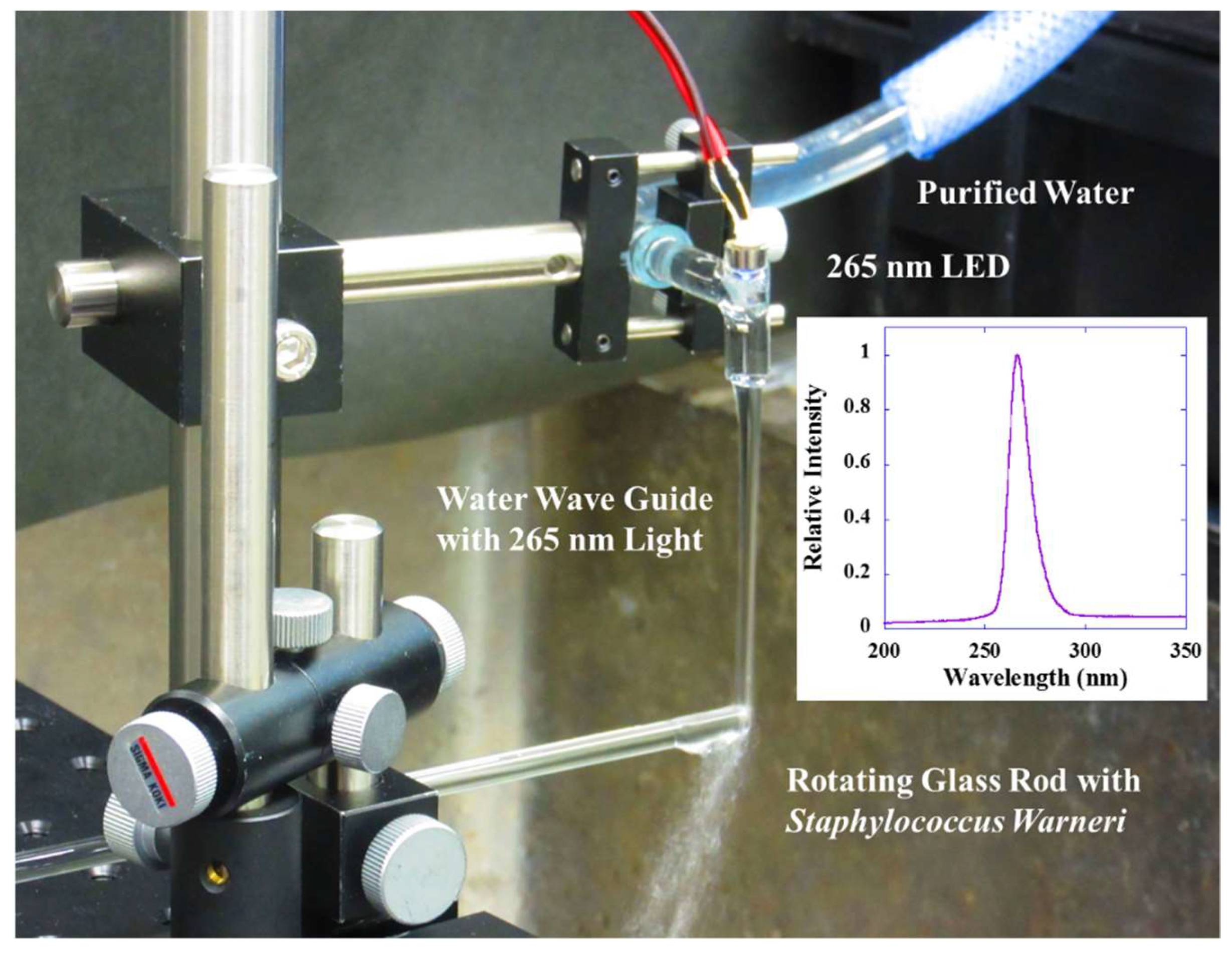
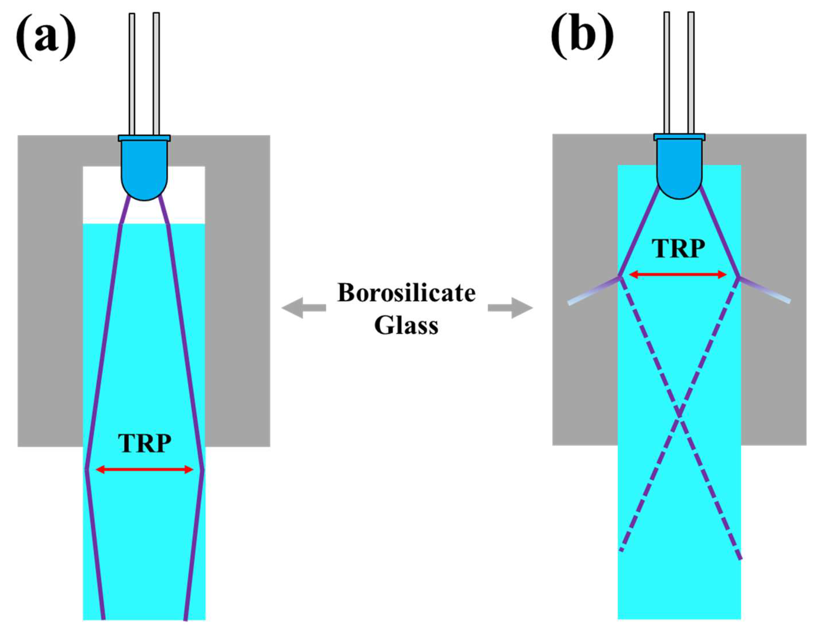
2.2. Theoretical Design and Experimental Setup for Water Waveguide System

The main components of the water waveguide disinfection system shown in Figure 1 are a TO-CAN type DUV-LED capped with a lens cap (VPT731 from Nikkiso Ltd. Tokyo, Japan), purified water supplied by a reverse osmosis (RO) system, and a rotating glass rod with a diameter of 5 mm. To perform the disinfections uniformly, the glass rod was rotated at 30 revolutions per minute during the disinfection tests (0–90 s). The emission wavelength of the DUV-LED was 265 nm with a spectral width (full width at half maximum) of 12 nm as measured by a spectrometer through an optical fiber (BIM-6002A, Brolight Technology Corporation, Hangzhou, China). The emission intensity of the LED was 0.5 mW at a forward voltage of 6 V and a driving current of 30 mA. The viewing angle of the 265 nm emission was determined by the DUV power as a function of distance between the LED and the irradiated area, and the power at the irradiated area was measured by a thermal surface-absorber-type power meter (PM16-401, Thorlabs Inc., Newton, NJ, USA). The viewing angle of the DUV emission was determined to be approximately 6°.
To introduce the 265-nm emission into the water stream, we used a T-shaped glass tube with an inner diameter of 6 mm. The flow rate of the purified water was maintained at 400 mL/min. Total reflection inside the water waveguide occurs at incident angles larger than 47° as estimated by Snell’s law; the refractive index of water, n, of 1.36 for this wavelength region was used for the estimation [44,45,46]. Therefore, DUV-LEDs with viewing angles smaller than 120° can be used for the water-waveguide method. Here, we note that LEDs generally have randomly polarized emissions; therefore, the reflectance at the air-water interface becomes larger with increasing incident angle. However, due to the small value of the refractive index of water, only 6.5% of the light is reflected when the incident angle is 60°. Therefore, efficient optical coupling between the DUV-LEDs and the water waveguides can be achieved without difficulty. Another advantage of using the water waveguide is the small extinction coefficient (κ) of water, κ = 2.75 × 10⁻⁸, which corresponds to a small absorption coefficient α of 0.013 cm−1 at an emission wavelength of 265 nm [44,45,46]. Therefore, almost all of the DUV emission can be guided; for example, theoretically, 85% of the DUV emission can be introduced to a disinfected glass rod 10 cm from the source.

3. Results and Analysis

3.1. DUV Fluence Measurements

The spectral intensity of 265 nm-DUV radiation at the head of the glass rod was measured by setting an optical fiber at the same place of the head and then the spectral intensity with or without the water stream was evaluated through the optical fiber and a spectrometer (BIM-6002A, Brolight Technology Corporation, Hangzhou, China). The DUV radiation power estimated by the intensity of the spectrum was 57 μW/cm2 without the water waveguide and 143 μW/cm2 with the water waveguide. The DUV radiation could be guided to a distant washing point (10 cm from the DUV-LED) with total internal reflection of the water stream; therefore, we could direct a 2–3 times higher DUV dose to the distant point compared to what could be achieved with simple DUV irradiation with the same disinfection time. The enhancement of the DUV light intensity due to internal reflection in the water flow is demonstrated by illuminating a phosphor tape (680MSH, 3M Japan Ltd., Tokyo, China) as shown in Figure 2.

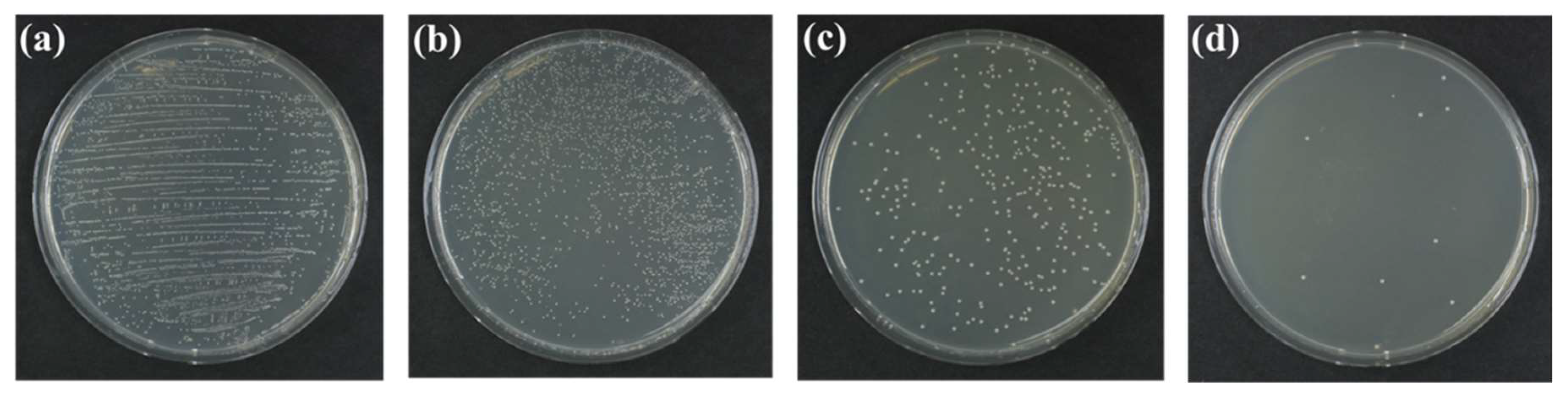
3.2. Efficacy of Disinfection by the Water-Waveguide Method

To quantitatively investigate the disinfection rates of the water-waveguide DUV disinfection system, we performed three experiments: (i) disinfection with DUV light, (ii) disinfection with running water, and (iii) disinfection with the water-waveguide method. For each experiment, the head of a glass rod (5ϕ × 270 mm) was dipped in a suspension of S. warneri, and then, the head of the glass rod was disinfected by one of the above three methods. To perform the disinfections uniformly, the glass rod was rotated at 30 revolutions per minute during the disinfection tests (0–90 s).
Figure 3 shows the results of (a) the control (before the disinfections), (b) disinfecting with DUV light (57 μW/cm2), (c) disinfecting with running water (400 mL/min), and (d) disinfecting with the water-waveguide method (143 μW/cm2 and 400 mL/min), where (b), (c), and (d) were performed for 1 min. The numbers of colonies counted were (a) 18,300 ± 4831 CFU for the control plate, reduced to (b) 3510 ± 1588 CFU by 1 min of DUV disinfection treatment, (c) 201 ± 141 CFU by 1 min of disinfection with running water, and (d) 9.3 ± 5.2 CFU by 1 min of water waveguide disinfection treatment. Here, the numbers reported are the means and standard deviations of CFU; dilutions (10−2–10−1) of the suspensions for (a) and (b) were made for accurate counting. In particular, statistically significant differences were clearly observed between (c) and (d), and its probability value (P) was calculated as P = 0.034.

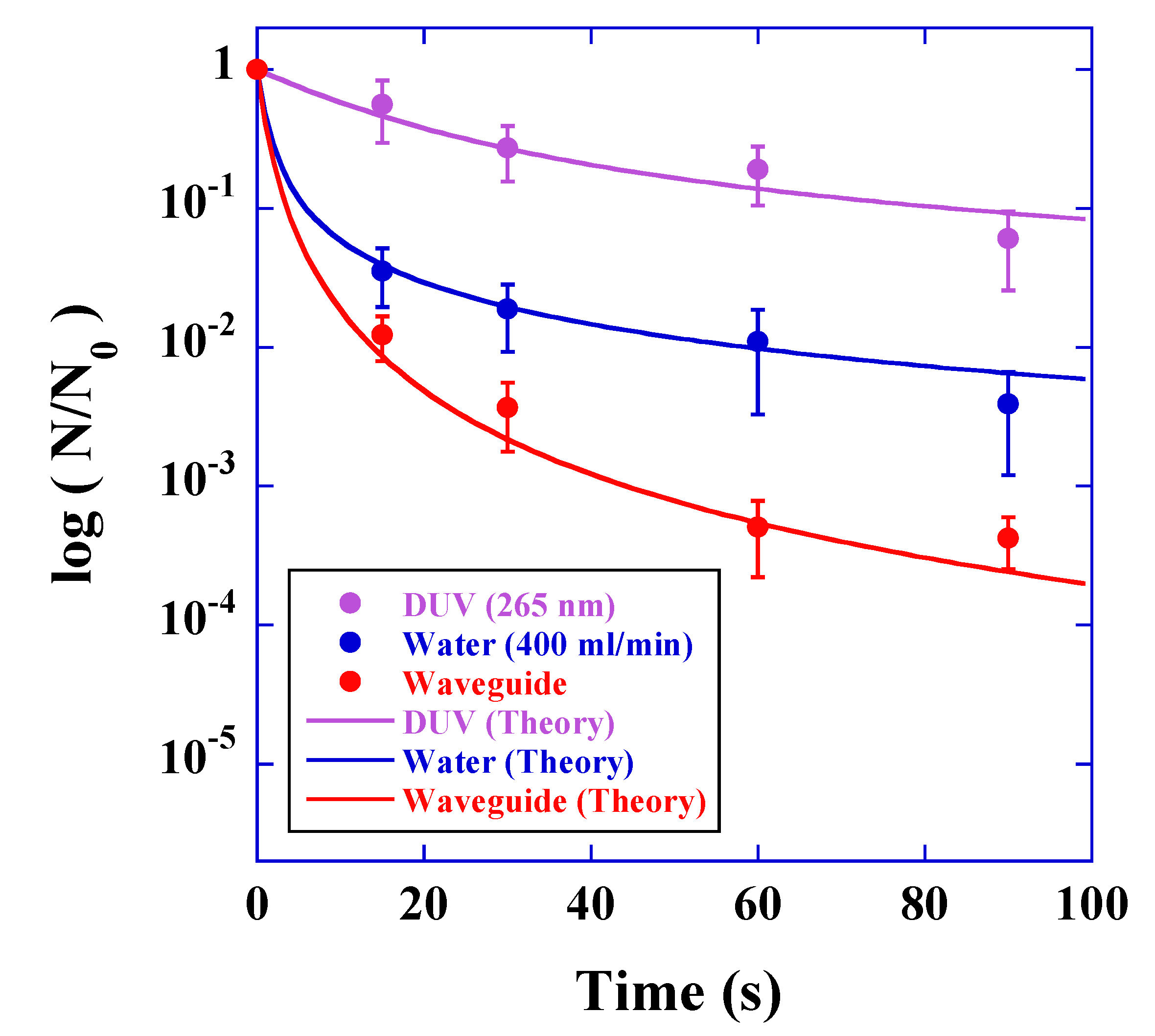
3.3. Theoretical Analysis of the Disinfection Rates

To quantitatively investigate the reduction of the disinfection rates as a function of duration, we plotted the relative ratio of the decrease in CFU (disinfected-CFU divided by control-CFU) caused by DUV light irradiation with an intensity of 57 μW/cm2 (purple circles), disinfection by running water at a flow rate of 400 mL/min (blue circles), and water-waveguide treatment with an intensity of 143 μW/cm2 and a flow rate of 400 mL/min (red circles), as shown in Figure 4. The reduction of the disinfection rates suggests that the decrease in CFU is not fitted by a mono-exponential reduction curve but is fitted by a multi-exponential curve (superposition of the mono-exponential reduction curve). It is evident that the multi-exponential reduction originates from variation between the environment of bacteria, because the DUV irradiation intensity and the adsorption strength that the bacteria sense are different from the position of the bacteria in the contaminated layer on the glass rod. These physical inhomogeneities affect the disinfection rates obtained by DUV light and/or running water. Thus, the relative rate of CFU reduction by the DUV irradiation, which is derived from the single-target model [47,48], can be expressed by the sum of the mono-exponential reduction curves with different reduction rate as
N D ( t ) N 0 = 1 exp ( Γ D I t ) Γ D I t
where N0 is the number of initial CFU; ND(t) is the number of CFU obtained by the DUV irradiation; t is the duration of the disinfection process; ΓD (cm2/mJ) is the photoinactivation rate, which depends on the environment of the bacteria and the irradiation wavelength; and I is the irradiation intensity of DUV light. Here we assume that the distribution of the reduction rates is uniform for the above calculation. To fit our results of the relative reduction rate of CFU by DUV irradiation, we used ΓD = 2.10 cm2/mJ, and I = 57 μW/cm2. The theoretically derived reduction rate described by Equation (1) is shown as a solid purple line. We note here that the value of ΓD obtained here is about three times smaller than the value previously reported [49]. We consider that this difference orginates from the shadow region (back side of the glass rod) when the glass rod is irradiated by the DUV radiation.
The theoretical rate of CFU disinfection with running water can be analyzed based on the kinetics of surface cleaning [50,51,52]. By taking the inhomogeneity in the disinfection process of running water into account, we find that the theoretical rate can be expressed as
N W ( t ) N 0 = 1 exp ( Γ W J t ) Γ W J t
where NW(t) is the number of CFU obtained by the running water treatment; ΓW (cm−1) is the disinfection rate of bacteria adsorbed on the surface of the glass rod, and J (cm/s) is the velocity of the water flow. To fit the results of infectivity by the disinfection with running water, we used the values J = 34.0 cm/s and ΓW = 0.05 cm−1. The theoretically derived relative rate of CFU obtained by these values is shown as a solid blue line.
The combined relative rate of the decrease in CFU by the water waveguide treatment can be expressed by the convolution function both of the photochemical disinfection and the running water given by Equations (1) and (2) as
N D W ( t ) N 0 = ( 1 exp ( Γ D I t ) Γ D I t ) ( 1 exp ( Γ W J t ) Γ W J t )
where NDW(t) is the number of CFU obtained by the water waveguide treatment. Here we used I = 143 μW/cm2 as the intensity of the DUV light. The same values, except this DUV intensity, were used for the fitting. The theoretically derived relative rate of the decrease in CFU obtained from these values is shown as a solid red line. The fitted curves agree well with the experimental results, and this agreement demonstrates that the water-waveguide is an efficient disinfection method that combines both physical disinfection with running water and photochemical inactivation.

4. Discussion

The time requirements for 1-Log (10−1) to 4-Log (10−4) reduction levels by DUV light, running water, and water-waveguide methods obtained by the experimental setup (shown in Figure 1) were theoretically estimated based on the experimentally determined parameters of ΓD and ΓW. The calculated results are presented in Table 1. The results show that higher reduction levels can be realized in a short period of time by using the water-waveguide method. For example, in order to obtain 3-Log level reduction, the duration of 590 s is required for running water treatment; however, it can be shortened to 44 s by using the water-waveguide method, leading to save 90% water consumption. More water consumption can be saved for higher Log reduction levels by using the water-waveguide method. Therefore, the water-waveguide treatment is a promising way for obtaining both a higher reduction levels and less water consumption.
Here we note that special care should be taken so that the lens of the DUV-LED is not immersed in water. This is because the T-shaped glass tube for obtaining the water waveguide effect is made of borosilicate glass material whose absorption coefficient at 265 nm wavelength region is large, more than 10 cm−1 [53,54]. Figure 5a shows that when the lens is not immersed in water, the DUV emission with incident angle of 3° in air refracts to 2° in water. The first total reflection point (TRP) can be taken to a distant position (66 mm in our case), and there is no absorption material at this position, hence the efficient total reflection occurs. However, as shown in Figure 5b, when the lens is immersed in water, the viewing angle of DUV emission becomes wider (30°) and the TRP moves closer to DUV-LED (4.3 mm in our case). The reflection occurs at the interface between water and the borosilicate glass. In this case, almost all the DUV emission transmits to the glass side (98%), where the DUV emission is almost absorbed due to the large absorption coefficient of the glass at 265 nm wavelength region. Therefore, we cannot obtain the DUV light waveguide effect with the water stream when the lens is immersed in water.
We have presented a new cleaning technique involving the use of water flow as the waveguide of DUV emission. The developed technique can be applied to various point-of-use treatments that can be installed in small spaces due to the small size of a DUV-LED, such as the disinfection of drinking water from taps. However, when we consider disinfecting drinking water, the water must be disinfected quickly (the time it takes for the water to move from the emission point of the DUV-LED to the end of the water stream (water waveguide region)). Therefore, a much higher intensity of DUV light is required. For example, assuming that the water flow rate is 100 mL/s, the water waveguide region is 10 cm, and the DUV light intensity in the water waveguide is constant, to inactivate Cryptosporidium parvum oocysts, the dose required for 3-Log inactivation is approximately 10 mJ/cm2 [1,5,20,55]. In this case, the required output power of the DUV light would be 85 mW at an emission wavelength of 265 nm (we determined these values based on the germicidal effectiveness curve [1,3,30]). These values can currently be achieved by using a high-power DUV-LED with a much wider spatial distribution and a viewing angle on the order of 120°. Total reflection inside the water waveguide occurs at angles larger than 47°, which corresponds to an incident angle of DUV-LED emission from air to water being approximately 60°; at this incident angle, reflectance loss is estimated as only 6%. Therefore, 94% of the DUV emission can be coupled to the water waveguide.
The water waveguide does not have a perfect smooth surface but a fluctuating shape; therefore, the condition of the total internal reflection is sometimes broken, leading to occur weak scattering of the DUV emission from inside to outside of the waveguide. Furthermore, the bacteria itself becomes the DUV scattering source. We have to take care of this leakage of the DUV emission, which would not exceed the threshold limit level (TLV) for UVC exposure. For example, in the United States of America, the TLV is 6 mJ/cm2 over an 8-hour period [1,56]. However, this DUV scattering can be protected by using commonly available transparent thin films such as polyethylene terephthalate (PET), because these films have large absorption coefficients at this DUV wavelength region.

5. Conclusions

We have successfully demonstrated a new water waveguide disinfection technique involving the use of a small, deep-ultraviolet light-emitting diode. The proposed water waveguide disinfection technique can be applied to many fields, such as washing techniques of skin, intraoral and internal-organ without the use of chemicals. The combination of the new technique demonstrated here powered by a small solar cell can provide a new convenient faucet for developing countries, which is possible to achieve efficient inactivation of a contaminated surface with less water consumption.

Author Contributions

T.M. is the co-first author. T.M., R.K. and T.F. contributed to the design of the water waveguide disinfecting system and completed all the disinfecting experiments. I.T. and T.H. provided technical support and bacterial expertise for the bacterial growth and colony-forming experiments. T.M., I.T. and T.H. constructed the quantitative model of the disinfection results. R.K and T.F. performed the statistical analysis for the infectivity experiments. All authors read and approved this submitted manuscript.

Funding

This research received no external funding.

Acknowledgments

The authors are grateful to Nikkiso Giken Corporation Ltd. for providing the DUV-LED devices and technical support for this study. The authors would like to thank Yuji Kohmura at Lucir Corporation Ltd. for his help with the hardware for rotating the glass rods. The authors would also like to thank Motoaki Iwaya at Meijyo University for information about the present status of DUV-LEDs.

Conflicts of Interest

The authors declare no competing interests.

References

  1. Pirnie, M.; Linden, K.G.; Malley, J.P. Ultraviolet Disinfection Guidance Manual for the Final Long Term 2 Enhanced Surface Water Treatment Rule; EPA 815-R-06-007; U.S. Environmental Protection Agency, Office of Water: Washington, DC, USA, 2006.
  2. Crawford, M.H.; Banas, M.A.; Ross, M.P.; Ruby, D.S.; Nelson, J.S.; Boucher, R.; Allerman, A.A. Final LDRD Report: Ultraviolet Water Purification Systems for Rural Environments and Mobile Applications; SAND2005-7245; Sandia National Laboratories: Albuquerque, NM, USA, 2005.
  3. Meulemans, C.C.E. The basic principles of UV–disinfection of water. Ozone Sci. Eng. 1987, 9, 299–313. [Google Scholar] [CrossRef]
  4. Bilenko, Y.; Shturm, I.; Bilenko, O.; Shatalov, M.; Gaska, R. New UV Technology for point-of-use water disinfection. In Proceedings of the 2010 Clean Technology Conference, Anaheim, CA, USA, 21–24 June 2010; pp. 339–342. [Google Scholar]
  5. Clancy, J.L.; Bukhari, B.; Hargy, T.M.; Bolton, J.R.; Dussert, B.W.; Marshall, M.M. Using UV to inactivate Cryptosporidium. J. Am. Water Work. Assoc. 2000, 92, 97–104. [Google Scholar] [CrossRef]
  6. Lindenauer, K.G.; Darby, J.L. Ultraviolet disinfection of wastewater: Effect of dose on subsequent photoreactivation. Water Res. 1994, 28, 807–814. [Google Scholar] [CrossRef]
  7. Ho, C.F.H.; Pitt, P.; Mamais, D.; Chiu, C.; Jolis, D. Evaluation of UV disinfection systems for large-scale secondary effluent. Water Environ. Res. 1998, 70, 1142–1150. [Google Scholar] [CrossRef]
  8. Linden, K.G.; Shin, G.A.; Sobsey, M.D. Comparative effectiveness of UV wavelength for the inactivation of Cryptosporidium parvum in water. In Proceedings of the 1st World Water Congress of the International Water Association, Paris, France, 3–7 July 2000; pp. 99–100. [Google Scholar]
  9. Oppenheimer, J.A.; Jacangelo, J.G.; Laine, J.M.; Hoagland, J.E. Testing the equivalency of ultraviolet light and chlorine for disinfection of wastewater to reclamation standards. Water Environ. Res. 1997, 69, 14–24. [Google Scholar] [CrossRef]
  10. Gray, N.F. Ultraviolet Disinfection. In Microbiology of Waterborne Diseases Microbiological Aspects and Risks; Academic Press: Waltham, MA, USA, 2014; pp. 617–630. [Google Scholar]
  11. Havill, N.L.; Moore, B.A.; Boyce, J.M. Comparison of the microbiological efficacy of hydrogen peroxide vapor and ultraviolet light process for room decontamination. Infect. Control Hosp. Epidemiol. 2012, 33, 508–512. [Google Scholar] [CrossRef] [PubMed]
  12. Anderson, D.J.; Gergen, M.F.; Smather, E.; Sexton, D.J.; Chen, L.F.; Weber, D.J.; Rutala, W.A. Decontamination of targeted pathogens from patient rooms using automated ultraviolet-c-emitting device. Infect. Control Hosp. Epidemiol. 2013, 34, 466–471. [Google Scholar] [CrossRef] [PubMed]
  13. Nerandzic, M.M.; Cadnum, J.L.; Eckart, K.E.; Donskey, C.J. Evaluation of a hand-held far-ultraviolet radiation device for decontamination of Clostridium difficile and other healthcare-associated pathogens. BMC Infect. Dis. 2012, 12, 120–125. [Google Scholar] [CrossRef]
  14. Nerandzic, M.M.; Thota, P.; Sankar, C.T.; Jencson, A.; Cadnum, J.L.; Ray, A.J.; Salata, R.A.; Watkins, R.R.; Donskey, C.J. Evaluation of a pulsed xenon ultraviolet disinfection system for reduction of healthcare associated pathogens in hospital rooms. Infect. Control Hosp. Epidemiol. 2015, 36, 192–197. [Google Scholar] [CrossRef]
  15. Haddad, L.E.; Ghantoji, S.S.; Stibich, M.; Fleming, J.B.; Segal, C.; Ware, K.M.; Chemaly, R.F. Evaluation of a pulsed xenon ultraviolet disinfection system to decrease bacterial contamination in operating rooms. BMC Infect. Dis. 2017, 17, 672–676. [Google Scholar] [CrossRef]
  16. Harm, W. Biological Effects of Ultraviolet Irradiation; Cambridge University Press: Cambridge, UK, 1980. [Google Scholar]
  17. Jagger, J. Yearly Review: Near-UV radiation effects on microorganisms. Photochem. Photobiol. 1981, 34, 761–768. [Google Scholar] [CrossRef] [PubMed]
  18. Jagger, J. Physiological effects of near-UV radiation effects on bacteria. Photochem. Photobiol. Rev. 1983, 7, 2–65. [Google Scholar]
  19. Friedberg, E.C.; Walker, G.C.; Siede, W. DNA Repair and Mutagenesis; ASM Press: Washington, DC, USA, 2006; pp. 92–107. [Google Scholar]
  20. Morita, S.; Namikoshi, A.; Hirata, T.; Oguma, K.; Katayama, H.; Ohgaki, S.; Motoyama, N.; Fujiwara, M. Efficacy of UV irradiation in inactivating Cryptosporidium parvum oocysts. Appl. Environ. Microbiol. 2002, 68, 5387–5393. [Google Scholar] [CrossRef] [PubMed]
  21. Reynolds, R.J.; Friedberg, E.C. Molecular mechanisms of pyrimidine dimer excision in Saccharomyces cerevisiae: Incision of ultraviolet-irradiated deoxyribonucleic acid in vivo. J. Bacteriol. 1981, 146, 692–704. [Google Scholar] [PubMed]
  22. Harris, G.D.; Adams, V.D.; Sorensen, D.L.; Curtis, M.S. Ultraviolet inactivation of selected bacteria and viruses with photoreactivation of the bacteria. Water Res. 1987, 21, 687–692. [Google Scholar] [CrossRef]
  23. Setlow, J.K. The effects of ultraviolet radiation and photoreactivation. Compr. Biochem. 1967, 27, 157–209. [Google Scholar]
  24. Beaven, G.H.; Holiday, E.R.; Johnson, E.A. Optical properties of nucleic acids and their components. In The Nucleic Acids; Chargaff, E., Davidson, J.N., Eds.; Academic Press: New York, NY, USA, 1954; Volume 1, pp. 493–553. [Google Scholar]
  25. Hall, J.D.; Mount, D.W. Mechanisms of DNA replication and mutagenesis in ultraviolet-irradiated bacteria and mammalian cells. Prog. Nucleic Acid Res. Mol. Biol. 1981, 25, 53–126. [Google Scholar]
  26. Sutherland, B.M.; Shih, A.G. Quantitation of pyrimidine dimer contents of nonradioactive deoxyribonucleic acid by electrophoresis in alkaline agarose gels. Biochemistry 1983, 22, 745–749. [Google Scholar] [CrossRef]
  27. Pfeifer, G.P. Formation and processing of UV photoproducts: Effects of DNA sequence and chromatin environment. Photochem. Photobiol. 1997, 65, 270–283. [Google Scholar] [CrossRef]
  28. Sinha, R.P.; Häder, D.P. UV-induced DNA damage and repair: A review. Photochem. Photobiol. Sci. 2002, 1, 225–236. [Google Scholar] [CrossRef]
  29. Cavaluzzi, M.J.; Borer, P.N. Revised UV extinction coefficients for nucleoside-5′-monophosphates and unpaired DNA and RNA. Nucleic Acids Res. 2004, 32, 1–9. [Google Scholar] [CrossRef] [PubMed]
  30. Beck, S.E.; Wright, H.B.; Hargy, T.M.; Larason, T.C.; Linden, K.G. Action spectra for validation of pathogen disinfection in medium-pressure ultraviolet (UV) systems. Water Res. 2015, 70, 27–37. [Google Scholar] [CrossRef] [PubMed]
  31. Song, K.; Mohseni, M.; Taghipour, F. Application of ultraviolet light-emitting diodes (UV-LEDs) for water disinfection: A review. Water Res. 2016, 94, 341–349. [Google Scholar] [CrossRef] [PubMed]
  32. Taniyasu, Y.; Kasu, M.; Makimoto, M. An aluminium nitride light-emitting diode with a wavelength of 210 nanometres. Nature 2006, 441, 325–328. [Google Scholar] [CrossRef] [PubMed]
  33. Khan, A.; Balakrishnan, K.; Katona, T. Ultraviolet light-emitting diodes based on group three nitrides. Nat. Photonics 2008, 2, 77–84. [Google Scholar] [CrossRef]
  34. Inoue, S.; Naoki, T.; Kinoshita, T.; Obata, T.; Yanagi, H. Light extraction enhancement of 265 nm deepultraviolet light-emitting diodes with over 90 mW output power via an AlN hybrid nanostructure. Appl. Phys. Lett. 2015, 106, 131104. [Google Scholar] [CrossRef]
  35. Takano, T.; Mino, T.; Sakai, J.; Noguchi, N.; Tsubaki, K.; Hirayama, H. Deep-ultraviolet light-emitting diodes with external quantum efficiency higher than 20% at 275 nm achieved by improving light-extraction efficiency. Appl. Phys. Express 2017, 10, 031002. [Google Scholar] [CrossRef]
  36. Inoue, S.; Tamari, N.; Taniguchi, M. 150 mW deep-ultraviolet light-emitting diodes with large-area AlN nanophotonic light-extraction structure emitting at 265nm. Appl. Phys. Lett. 2017, 110, 141106. [Google Scholar] [CrossRef]
  37. Messina, G.; Burgassi, S.; Messina, D.; Montagnani, V.; Cevenini, G. A new UV-LED device for automatic disinfection of stethoscope membranes. Am. J. Infect. Control 2015, 43, e61–e66. [Google Scholar] [CrossRef]
  38. Messina, G.; Fattorini, M.; Nante, N.; Rosadini, D.; Serafini, A.; Tani, M.; Cevenini, G. Time effectiveness of ultraviolet C light (UVC) emitted by light emitting diodes (LEDs) in reducing stethoscope contamination. Int. J. Environ. Res. Public Health 2016, 13, 940. [Google Scholar] [CrossRef]
  39. Chevremont, A.C.; Farnet, A.M.; Coulomb, B.; Boudenne, J.L. Effect of coupled UV-A and UV-C LEDs on both microbiological and chemical pollution of urban wastewaters. Sci. Total Environ. 2012, 426, 304–310. [Google Scholar] [CrossRef] [PubMed]
  40. Beck, S.E.; Ryu, H.; Boczek, L.A.; Cashdollar, J.L.; Jeanis, K.M.; Rosenblum, J.S.; Lawal, O.R.; Linden, K.G. Evaluating UV-C LED disinfection performance and investigating potential dual-wavelength synergy. Water Res. 2017, 109, 207–216. [Google Scholar] [CrossRef] [PubMed]
  41. Li, G.; Wang, W.; Huo, Z.; Lu, Y.; Hu, H. Comparison of UV-LED and low pressure UV for water disinfection: Photoreactivation and dark repair of Escherichia coli. Water Res. 2017, 126, 134–143. [Google Scholar] [CrossRef] [PubMed]
  42. Sutton, R.M. Demonstration Experiments in Physics; McGraw-Hill: New York, NY, USA, 1938; Volume 1, p. 385. [Google Scholar]
  43. Kshatriya, A. Water jet as a laser pipe. Am. J. Phys. 1976, 44, 604. [Google Scholar] [CrossRef]
  44. Hale, G.M.; Querry, M.R. Optical constants of water in the 200-nm to 200-µm wavelength region. Appl. Opt. 1973, 12, 555–563. [Google Scholar] [CrossRef] [PubMed]
  45. Daimon, M.; Masumura, A. Measurement of the refractive index of distilled water from the near-infrared region to the ultraviolet region. Appl. Opt. 2007, 46, 3811–3820. [Google Scholar] [CrossRef] [PubMed]
  46. Quickenden, T.I.; Irvin, J.A. The ultraviolet absorption spectrum of liquid water. J. Chem. Phys. 1980, 72, 4416–4428. [Google Scholar] [CrossRef]
  47. Atwood, K.C.; Norman, A. On the interpretation of multi-hit survival curves. Proc. Natl. Acad. Sci. USA 1949, 35, 696–709. [Google Scholar] [CrossRef] [PubMed]
  48. Meynell, C.G.; Meynell, E. Theory and Practice in Experimental Bacteriology, 2nd ed.; Cambridge University Press: Cambridge, UK, 1970; ISBN 978-0-521-07682-1. [Google Scholar]
  49. Shoults, D.C.; Ashbolt, N.J. Decreased efficacy of UV inactivation of Staphylococcus aureus after multiple exposure and growth cycles. Int. J. Hyg. Environ. Health 2019, 222, 111–116. [Google Scholar] [CrossRef]
  50. Jennings, W.G. Theory and practice of hard-surface cleaning. Adv. Food Res. 1965, 14, 325–458. [Google Scholar]
  51. Levenspiel, O. Chemical Reaction Engineering, 3rd ed.; John Wiley & Sons: New York, NY, USA, 1999; ISBN 0-471-25424-X. [Google Scholar]
  52. Cutler, W.G.; Kissa, E. Detergent: Theory and Technology; Marcel Dekker, Inc.: New York, NY, USA, 1987; ISBN 0-8247-7503-1. [Google Scholar]
  53. Kitamura, R.; Pilon, L.; Jonasz, M. Optical constants of silica glass from extreme ultraviolet to far infrared at near room temperature. Appl. Opt. 2007, 46, 8118–8133. [Google Scholar] [CrossRef] [PubMed]
  54. Refractive Index Database. Available online: https://refractiveindex.info/ (accessed on 10 December 2018).
  55. Shin, G.A.; Linden, K.G.; Arrowood, M.J.; Sobsey, M.D. Low-Pressure UV inactivation and DNA repair potential of Cryptosporidium parvum oocysts. Appl. Environ. Microbiol. 2001, 67, 3029–3032. [Google Scholar] [CrossRef] [PubMed]
  56. Nardell, E.A.; Bucher, S.J.; Brickner, P.W.; Wang, C.; Vincent, R.L.; Becan-McBride, K.; James, M.A.; Michael, M.; Wright, J.D. Safety of upper-room ultraviolet germicidal air disinfection for room occupants: Results from the tuberculosis ultraviolet shelter study. Public Health Rep. 2008, 123, 52–60. [Google Scholar] [CrossRef] [PubMed]
Figure 1. Water waveguide disinfection system. To introduce the 265-nm emission into the water stream, a T-shaped glass tube with an inner diameter of 6 mm was used. The emission wavelength of the deep-ultraviolet light-emitting diode (DUV-LED) was 265 nm, and its spectral width was 12 nm, as shown in the inset spectrum. Purified water supplied from a reverse osmosis system with a flow rate of 400 mL/min was used for the disinfection experiments. To perform the disinfection uniformly, each glass rod (after dipping the head in the bacterial suspension) was rotated at a speed of 30 revolutions per minute during the disinfection tests.
Water 11 00294 g001
Figure 2. DUV fluence of the water-waveguide effect. The radiation power estimated by the intensity of the spectrum was (a) 57 μW/cm2 for DUV radiation without the water-waveguide effect and (b) 143 μW/cm2 for DUV irradiation with the water-waveguide effect. The enhancement of the DUV light intensity achieved with the water-waveguide effect is demonstrated by illuminating a phosphor tape.
Water 11 00294 g002
Figure 3. Results of disinfection for S. warneri; (a) control, (b) disinfecting by DUV light (57 μW/cm2), (c) disinfecting by running water (400 mL/min), and (d) disinfecting by the water-waveguide method (143 μW/cm2 and 400 mL/min); the durations of treatments (bd) were 1 min. The number of colonies counted were (a) 18,300 ± 4831 colony forming unit (CFU) for the control plate, reduced to (b) 3510 ± 1588 CFU by 1 min of DUV disinfection treatment, (c) 201 ± 141 CFU by 1 min of the disinfection with running water, and (d) 9.3 ± 5.2 CFU by 1 min of water-waveguide treatment. Here, the numbers of CFU are reported as the means and standard deviations of at least three independent experiments. For counting the number of CFU of original (a,b) plates, dilutions (10−2–10−1) of the suspensions for (a,b) were prepared. Significant differences were observed between (c,d), and the probability value (P) was found to be P = 0.034.
Water 11 00294 g003
Figure 4. The relative ratio of the decrease in CFU for S. warneri (disinfected-CFU divided by control-CFU) achieved with DUV light irradiation with an intensity of 57 μW/cm2 (purple circles), running water treatment with a flow rate of 400 mL/min (blue circles), and water-waveguide treatment with an intensity of 143 μW/cm2 and a flow rate of 400 mL/min (red circles). The theoretically calculated lines express the rate of the decrease in CFU by DUV irradiation (purple line), running water treatment (blue line), and water-waveguide treatment (red line).
Water 11 00294 g004
Figure 5. Change of the total reflection point (TRP) when the head of the LED-lens is (a) not immersed in water or (b) immersed in water. (a) When the lens is not immersed in water, the DUV emission with incident angle of 3° in air refracts to 2° in water. The first TRP point can be taken to a distant position (66 mm in our case), and there is no absorption material (borosilicate glass) at this position, thus the efficient total reflection occurs. (b) When the lens is immersed in water, the viewing angle of the DUV emission from the lens becomes wider (30°) and the TRP moves closer to the DUV-LED (4.3 mm in our case). The 98% transmission of the DUV emission occurs from water to the borosilicate glass and in this case we cannot obtain the water-waveguide effect of the DUV light.
Water 11 00294 g005
Table 1. Time requirements to obtain the 1-Log to 4-Log reductions by DUV light, running water, and water-waveguide disinfection methods for S. warneri. The unit used here is seconds.
Disinfection MethodsLog(N/N0) = −1Log(N/N0) = −2Log(N/N0) = −3Log(N/N0) = −4
DUV Irradiation8.3 × 1018.3 × 1028.3 × 1038.3 × 104
Running Water5.95.9 × 1015.9 × 1025.9 × 103
Water Waveguide3.61.4 × 1014.4 × 1011.4 × 102

